Michael Beug
Introduction to Jelly fungi, by Robert Bandoni
Techniques in microscopy of Jelly fungi, by Robert Bandoni
Purple or pink or pale lavender
Colorless (hyaline) to white or gray
The term "jelly fungus" covers a large variety of fungi that share a characteristic textural feature of having gelatinous sporocarps, i.e. the hyphae and reproductive structures are embedded in a gelatin-like matrix. Supposedly, the gelatinous material results from the breakdown of outer hyphal wall layers, but there is little evidence for this origin. What does appear to be the case, however, is that many of these fungi can dry down to an inconspicuous layer or forms, take up water, revive and sporulate. Furthermore, some can do this repeatedly. The delicate parts of the sporocarp (asci, basidia, other reproductive structures), are well-protected in the dried state, the very hard material then resistant to the activities of mites, insects, vertebrates, etc. In fact, basidiomes of some groups of jelly fungi, e.g., Merulius spp., have restricted portions of the sporocarp the hymenium and some subhymenial material that are gelatinous. Many of the taxa dealt with here, however, have sporocarps that are completely gelatinous. The major exception to this is that many Tremella spp., all of which are probably mycoparasites, have hard cores of host hyphae in their basidiocarps. And it is interesting to note that these cores belong to basidiomycetes some of which produce sporocarps which lack gelatinous layers but can survive for long periods in the dried condition.
The ability to dry, rehydrate and revive when small amounts of moisture become available seems to be most advantageous to many species growing on recently dead branches of woody plants. Dead branches in the canopy, or dead standing trees, are the most common habitats for many species. Windblown branches from the canopy often bear unusual jelly fungi, as do the dead, lowermost branches of many trees, both broad leaf and coniferous. Recently fallen trees and branches are also excellent substrates to examine for these fungi. Since many jelly fungi are mycoparasites, it is a good idea to examine sporocarps of ascomycetes and basidiomycetes on such trees. The basidiocarps of many host fungi, e.g. Stereum, Aleurodiscus, Diatrype, etc. can be much more conspicuous than their parasites. Knowing this, conspicuous potential host sporocarps can help locate the more inconspicuous parasitic species. Using this method often allows one to collect even dried basidiomes of larger jelly fungi that occur near or on their host sporocarps and cannot then be seen from a distance. It should be added that many basidiomycetous mycoparasites are reduced to hyphae and reproductive cells that develop within the sporocarp of the host, and these are seen in microscope preparations.
The jelly fungi discussed in these keys amount to a hodge-podge taxonomically: they are not a close- knit group. They include selected spp. of Tremellomycetidae, Urediniomycetidae and Discomycetes (Ascomycetes) plus a few Homobasidiomycetous forms in the Meruliaceae and several other families. Their individual lifestyles differ strongly as some are parasites of other fungi (including other jelly fungi), parasites of green plants, parasites of the symbiotic pairs in lichens, symbionts of higher plants in the form of mycorrhizae. Many are simply decay fungi (the most important activity carried on by fungi generally).
The keys are not based upon microscopy, but many taxa are essentially indistinguishable macroscopically. Consequently, microscopy must be used to finalize identification in all cases. The superficial characters used in the keys must include every item visible, i.e., not just of the basidiomes, but their habitat, consistently associated plants or animals, and substrates. A list follows.
Key Characters
Color: Often bright and distinctive, also inconsistent in many cases, changing with level of moisture, age, exposure in some cases, spore state present, and probably with associated fungi. Various color charts have been published, but no two individuals probably see color in the same way. Approximations generally are sufficient, since the color is variable. Color reference books, if available, allow one to compare named colors in a book of plates to the fungus on hand. This should be done with material in the fresh state, then noted also for the dried state.
Texture: Here, the texture is much like that of gelatin, quivery like Jello, but becoming tough, then hard upon drying. The gelatin-like layers may be associated with relatively fleshy ones in which the hyphae do not develop in a matrix, but remain more or less separate from one another.
Form and Size: Form, like color and texture, is generally variable. It is most consistent in some highly developed forms such as Phlogiotis, Pseudohydnum, several genera of Dacrymycetaceae, and others. For many Tremellales, the largest group species-wise, and, so far as is known at present, probably all mycoparasites, growth of the basidiome is indeterminate, continuing for as long as weather and the host permit. They then can dry, revive if wet again, the growth again is indeterminate and generally the form is irregular. Nor are size and form consistent in many other jelly fungi, as drying and revival of growth results in mixed sizes of sporocarps. Form also is altered by anastomosis of adjacent sporocarps, i.e., they grow together leaving a visible join or none, but causing a change in shape.
Measurements: These should be taken as soon as possible after collecting because they change rapidly as the sporocarps lose moisture. If necessary the measurements can be approximated by soaking a sporocarp after it has dried, but measuring the fresh sporocarp gives the best results.
Surface features:
Hymenophore: smooth, toothed, poroid, lamellate.
Non-fertile or fertile surfaces: smooth, scabrid, hairy, villose, warty, bullate, grooved.
Internal inclusions other than hyphae: pigment granules, accretions of lime.
Habitat and substrate: Many jelly fungi grow on wood. It should be noted whether the fungus is on a stump, on a fallen trunk, on a branch, on woody debris, or on a standing tree, whether living or dead, and whether the bark is still in place. The tree species should be determined when possible. Even if the tree species cannot be determined with certainty, it is helpful to know if the tree is a broad-leaved or coniferous species. Other fungi are mycoparasites (they grow on other fungi). Associated sporocarps of other basidiomycetes or ascomycetes (including lichens) should be noted.
Season: Jelly fungi, like other fungi, develop sporocarps more commonly in some seasons and weather conditions than others. Heterotextus alpinus, for example, occurs more often in late winter. Other species develop sporocarps more often in fall. By contrast, Exidia recisa is present over a longer period from spring until fall if weather is wet.
Spore print: Making a spore print with the fresh material requires only that a sporocarp be allowed to dry over night on a microscope slide or appropriate paper. The allows superimposing the spores over very white or black paper to determine shades of color; it also eliminates the need for scraping spores away from a paper print in order to make a microscope preparation.
Microscopic features:
A. Basidial morphology: There are numerous variations in basidial form in this group, as follows:
1) "Auricularioid" basidia. These are cylindric, transversely septate, and are 2-4 -celled when mature. Each cell produces a sterigma or a lateral extension bearing a spore. Typical of species of Auricularia, Platygloea, Stilbum, Atractiella, and relatives of these as well as in the major groups of rusts and many smut fungi.
2) "Tremelloid" basidia. These are globose to ellipsoidal or stalked and capitate, the swollen part vertically septate and 2-4 -spored, vertically partially septate or the septa variously arranged, i.e., part diagonal, or transverse. Some members of the group also have holobasidia; these may be clavate or long cylindrical.
3) Holobasidia. Non-septate, commonly club shaped and bearing two spores, e.g. as in the Dacrymycetaceous species ("tuning fork basidia"). Some Tremellales have holobasidia with 2 - 4 terminal spores.
B. Spore characteristics, including size and shape, color in mass, surface features (e.g., spines)
C. Basidiospore germination: Basidiospores can germinate in several ways and these are sometimes diagnostic. In the Tremellomycetidae and many Urediniomycetidae, the basidiospores germinate by budding, yielding unicellular budding cells (yeast states). But, in these same groups, spores can also germinate by forming mating tubes if compatible spores are present nearby. Many basidiospores of heterobasidiomycetes can also germinate by repetition. In this process, the spore produces a sterigma similar to those seen on many basidia; this is tipped by a basidiospore that usually resembles the parent spore and, like it, is shot from the sterigma. Various types of conidia (asexual spores) other than repetitive ones can develop from a germinating basidiospore. The types of such secondary spores produced by basidiospores upon germinating are taxonomically important.
D. Characteristics of hyphae including presence of clamp connections
E. Yeast states
Preparing Sections
Preparing microscope slides from fresh jelly fungus basidiocarps can be a challenging task but it need not be difficult if one has appropriate tools, procedures to follow, and mounting solutions. The tools needed are: a new single edge razor blade, a pair of dissecting needles, preferably with wooden handles, and elder pith or a substitute if you can obtain these. The needles can be sharpened in such a way as to produce a knife edge near the tip. To do this, first remove a bit of the wooden handle on two sides adjacent to the needle: the cuts should be at an angle to each other. This will allow you to use a medium coarse carborundum block to sharpen and shape the needle tip at an angle. Hold the needle by the handle, the index finger placed on one of the two cuts, make slow figure eights with the needle at a relatively flat angle to the carborundum surface and continue the motion until tip is ground off at a slight angle. Change the index finger to the other notch, repeat the motion. If done properly, the tip becomes knife like and is suitable for actually removing small pieces of fresh sporocarps. When finished sharpening the needle, clean the tip, place a fresh specimen on the stage of a dissecting microscope, remove a tiny bit of the hymenium or other desired portion of the specimen. Add this material to a drop of stain or mounting fluid on a microscope slide, allow to soften and stain for a few minutes, then cover with a cover slip. Excess fluid should be absorbed with paper toweling from the edge of the mount, at the same time applying slight pressure on the covers glass directly over the bit of material (use the needle handle or a similar tool, not your fingers!). If you are lucky, the fragment will flatten and you can examine it under high magnification with a compound microscope. Unfortunately, the sole result may also be that the cover glass shatters and the specimen does not flatten. This technique works well for soft material, e.g., gills of mushrooms, some soft gelatinous resupinate heterobasidiomycetes on wood, etc. With things such as Auricularia sporocarps, the "flesh" is extremely tough (and the cover glass is fragile!) Don't throw away the needles in disgust, however, as they are useful in other ways.
If you can either collect or buy pieces of elder pith(1), these are useful for cutting very thin sections of fresh sporocarps, or pieces of these (or dried ones that have been rehydrated in water). Commercial pith has all of the surrounding woody layers removed. A piece of pith 4-5 cm long can be made into a specimen holder by splitting it lengthwise into two equal halves. A portion of a sporocarp(2), or an entire sporocarp, can be placed on the flat side of one piece; the second is placed on top of the material and the two are grasped firmly between the thumb and forefinger of one hand (left hand for right handers, right hand for left handers). Using pressure to grip the material between he two halves of the pith, sections are then cut with the free hand from the end clasping the material. The first slice trims the excess material away; subsequent slices remove sections of the material (a section of pith also falling away each time a slice is made). With practice, holding the elbows in next to the body and slicing toward the chest, sections 20-30 µm thick are possible. The sections can be dropped into a shallow dish of water as they are sliced away from the pith. Under a dissecting microscope at 20 X, it is possible to select the best sections (thinnest ones, and those showing desired surfaces, etc.). Insert the needle under a section, lift and transfer it to a drop of mounting fluid on a microscope slide. It is not absolutely necessary to have a complete 10 µm section of the desired portion of the material. Frequently, the thin edges of wedge-shaped sections are excellent (these are more common than the perfectly level ones).
If you have some free time and a razor blade, but no pith, the following is recommended. Allow the specimen (or a portion of) it to dry until it is brittle. Place the fragment on the stage of a dissecting microscope, and focus to the best position. This should be done at relatively low magnification as one then grasps the single edge razor and, while viewing the material under the dissecting scope, slice sections from the edge of the dry material. With practice, it is possible to cut sections as thin as those done with pith (above) or possibly thinner. The sections are transferred directly to mounting fluid on a glass slide, allowed to rehydrate and stain for a minute, and a cover glass is then added to the preparation.
The commonest error made in preparing microscope slides is the addition of too much material (small, thin beats fat and thick every time). It is easier to prepare another slide, than to remove material from one that is overloaded. Use the thinnest slices you can obtain; flatten them further if needed, but do not apply too much pressure!!!
Measuring Spores
At least 10-20 spores should be measured to get a reasonable range for a species. Those present on the hymenium are often secondary ballistospores that are slightly larger than their parents - which need not be the first generation after the basidiospore(3). It's better to use spores in a spore print. (Although these do not differ greatly from those on the hymenium in either form or size, they are sometimes slightly larger from the print.) Prints should be made on glass slides.(4) 3% KOH may be used, or a drop of Lacto-phenol-Cotton Blue or Lacto-fuchsin can be added to one edge of the spore print to prepare spores for measuring(5). With glass microscope slides, the color if any can be seen more clearly by placing the slide on a paper background - white, black or whatever. These slides can be stored in boxes, but also can be put in cardboard "mailers" and mailed together with the specimen.
Stains and Mounting Fluids
Sections can be mounted in water, covered with a cover glass, and viewed immediately. However, special mounting fluids may be necessary to bring dry material back to its original state of turgor. The most commonly used fluid for this purpose is 3% KOH (3 gm per 100ml. distilled water), often obtainable from pharmacies. Also, for jelly fungi, this mounting fluid is commonly used with stain solutions, especially aqueous Phloxine 1% and or aqueous Congo Red 1%. Phloxine is a cytoplasmic stain; Congo Red, in alkaline solutions such as this, is an excellent wall stain. In preparing slides with either of these, a drop of each of the KOH and the desired stain are placed together on a glass slide; a section or sections are added, allowed to stain briefly, then covered. Excess fluid should be removed from the preparation before examining it. If slides are for special purposes, such as photography, it may be best to clear the slide of staining solution. To do this, hold a piece of absorbent toweling in contact with the liquid at one edge of the cover glass; at the same time, replace the stain-KOH mixture with a dropper at the opposite edge. Note that 3% KOH is rather caustic and can fog lenses; any that accidentally gets on lenses should be cleaned immediately.
Aqueous microscope preparations such as those above will dry out quickly, but additional stain or KOH can be added at the edge of the cover glass to prevent excessive drying. Alternatively, if one wishes to keep such preparations for years, 10% glycerol can be added at the edge of the cover slip as drying occurs. Continue to do this until evaporation appears to cease. The cover slip can be "glued" in place at that time by using nail polish. Do not attempt to "seal" the edges with nail polish or similar materials as the glycerol is slowly volatile (in spite of the seal) and drying generally occurs. By fixing only the corners of the cover glass to the slide, the spaces between these points can be used to add new fluid to the preparation. Slides of these types can be kept for 50 years or more, rehydrated, and are as "good as new".
Other mounting fluids commonly used are Melzer's Reagent and Lacto-Phenol-Cotton Blue. Melzer's reagent contains iodine and it will react with starch or starch-like substances to give a deep blue-black color (amyloid reaction). Some fungal structures turn reddish brown (dextrinoid reaction) in this reagent. Jelly fungi do not appear to have substances reacting with Melzers reagent, but this solution may still be used because of the clear picture it seems to give (which may be related to refractive index). Lacto-Phenol-Cotton Blue also acts as a chemical test reagent with fungal material and it does give a visible cyanophilous reaction with certain spore walls or some layers of spore walls. This reagent is also widely used as a mounting medium for many kinds of fungi as the preparations retain their usefulness for long periods (they do not dry quickly and some modern versions evaporate even less quickly). Both of the above mounting media can be difficult to obtain as they contain the toxins phenol (Lacto-Phenol-Cotton Blue), and chloral hydrate (Melzer's). Commercial grade lactic acid can also be used for a general purpose mounting fluid: it is usually mixed with a small amount of Acid Fuchsin dye. Finally, either dilute alkaline safranine or very dilute solution of Azure Blue B to see nuclei clearly. One drop of stain and one drop of KOH seems to work well.
(1) I have collected natural pith only once or twice - collecting the relatively thick first years growth of sucker shoots. They have only a thin layer of woody tissue and bark around the outside and it can be cut away. The pieces I got were neither as large as those from supply houses, nor were they as clean. It should have a consistency considerably more delicate than cork - very easily cut or sliced into section with a razor blade. It is extremely light. I have heard that you can use some of the types of foam plastics used in "peanuts" and the like, but have never tried this. I think they would be tougher than pith.
(2) The pith should be dry; if you wet it, you have troubles. It is best to remove all surface water from the portion of basidiocarp that you clamp between the pieces of pith.
(3) It is often difficult to know whether spores measured by many individuals come from the hymenium or from spore prints. I say hymenium here because generally only very small spores are found attached to the basidia. With rather primitive basidiomes such as in jelly fungi, basidia are mostly directed up, down, sideways, etc. Many such spores simply fall back on the hymenium and dry together with the sporocarp. Alternatively, they may give rise to secondary spores (via germination bye repetition); the repeats can also fall back onto the hymenium. These exposed surfaces are sometimes "adorned" with spores of several other types, not just those of the jelly fungi. So, one needs to know what to look for when measuring loose spores - there probably are errors at times, but not so many if one has some idea what to expect.
(4) Kenneth Wells likes to dry several viable specimens with the fertile surface up, and finds that this gives the best specimens. Even so so one sometimes has to do a lot of searching. Drying the specimens over dark paper will also supply masses of spores. The spores may be lifted off the paper in a drop of 3% KOH or water. Dr. Wells finds that if specimens are properly dried, spore measurements can be made indefinitely and do not seem to differ from those made of a fresh specimen. He measures spores in Phloxine and KOH or Phloxine, Congo Red, and KOH, and finds it then easy to see the spore walls for measurements. Ian Gibson.
(5) Spore measurements are not expected to differ substantially when made in water (fresh specimens) or in 3% KOH (fresh or dried specimens), but the stains make the spore walls more visible. Ian Gibson
I am grateful to Dr. Robert Bandoni for his help in assembling source materials, for his review of the draft key and suggested improvements, for his written contributions above, and also for his many years of research contributing to the literature on this group of fungi. Dr. Kenneth Wells, another prolific researcher on jelly fungi, was also very helpful in answering my questions.
The key includes those gelatinous fungi (basidiomycetes or ascomycetes) that reach at least 5 mm in diameter and 3 mm in height or thickness in their fresh state, whether as individual sporocarps or as aggregations of sporocarps. It is not intended to cover resupinate fungi (those forming a spreading paint-like layer on their substrate), of which a large number are considered jelly fungi either by their family connections or their gelatinous consistency. (Some of the excluded resupinate species, Exidiopsis plumbescens for example, start as small pustules, generally less than 5 mm in diameter. The pustules then enlarge, often anastomosing more or less completely although the surface in some species may show a distinct pattern of the original pustules.) A few very small Dacrymyces and Tremella species are included in this key for comparison. Some of these do not reach 3 mm in thickness and therefore would not strictly fit the criteria.
If the microscopic features are in parentheses, they are not always necessary if the species is familiar, but are helpful.
The species are divided in the key into five groups characterized by the color of fresh, moist sporocarps. Species that have more than one color are included in more than one color group. The key leads start at the following numbers.
Yellow to orange when fresh and moist
................................................................................101
Purple or pink or pale lavender when fresh and moist
................................................................................201
Pale green or yellowish green
................................................................................301
Yellow to orange when fresh and moist
................................................................................401
Black or smoky transparent when fresh and moist
................................................................................501
Colorless (= hyaline) to white or gray when fresh and moist
................................................................................601
In more detail, the colors are keyed in the following fashion. Colors refer to the fresh, moist state: colors in age or when dry are often darker, faded, or different.
100a Pale dull yellow to bright yellow, orange or sometimes reddish orange
................................................................................101
100b Not yellow to orange (may be white to very faintly yellowish, yellow-brown)
................................................................................200
200a Purple or pink or pale lavender when fresh and moist
................................................................................201
200b Not purple or pink or pale lavender (may be vinaceous, liver-brown, brownish flesh-colored, very pale pink)
................................................................................300
300a Pale green or yellowish green (may dry black)
................................................................................301
300b Not pale green or yellowish green
................................................................................400
400a Black or smoky transparent when fresh and moist
................................................................................401
400b Not black
................................................................................500
500a Colorless to white or gray (or pale amber or pale yellow or pale pink )
................................................................................501
500b Various shades of brown (not colorless to white or gray, may be gray-brown)
................................................................................601
The color of dried sporocarps can be helpful at times - for example in Tremella exigua the color of developing sporocarps is often whitish (pale "milky") with a greenish cast, but the dried sporocarps typically are greenish black. Some information about colors of dried sporocarps is presented in the descriptions.
YELLOW TO ORANGE
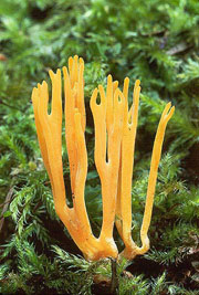
101a Erect yellowish coral-like mushroom, with antler-like branching, up to 10 cm high
................................................................................Calocera viscosa
Note: Ramaria species are coral-like but gelatinous species are larger with more profuse branching.
| BASIDIOCARP up to 10 cm high, expanded above a short stipe about 3 mm wide into erect, repeated, antler-like, dichotomous branches that are round in cross-section or flattened, often up to several fused together at the base; firm-gelatinous, slippery; golden yellow or orange-yellow. HABITAT on rotting conifer wood or, more commonly, growing beside rotten conifer stumps. MICROSTRUCTURES spores 8-12.5 x 3.5-4.5 um, cylindrical to slightly curved-cylindrical, becoming 1-septate at maturity; germination by colorless, globose to subglobose conidia or by germ tubes; basidia shaped like tuning forks; occasionally, simple cylindrical hyphidia (dikaryophyses) occur between basidia; clamp connections absent. REMARKS Coral fungi such as Ramaria either have soft fragile flesh or are gelatinous in parts but much larger, whereas Calocera viscosa is tough and difficult to tear. Clavulinopsis corniculata is similar and also somewhat tough, but is not viscid, often grows on the ground, and is different microscopically. These clavarioid fungi do not have the tuning fork type of basidia and their spores are not septate. | Calocera viscosa Michael Beug |
101b Not erect, or not coral-like, or not branching
................................................................................102
102a Erect basidiocarps that are cylindrical to awl-shaped and rarely forking
................................................................................Calocera cornea
| BASIDIOCARP up to 2 cm high, simple, awl-shaped, occasionally forked or palmate or short-stipitate with a folded or wrinkled head, firm-gelatinous, slippery; yellow to orange-yellow, drying red-brown. HABITAT on barkless branches of hardwoods, less often on conifer wood. MICROSTRUCTURES spores 7-10 x 2.5-4.5 um, cylindrical to slightly curved, becoming 1-septate at maturity; basidia finally the shape of a tuning fork; simple cylindrical hyphidia sometimes present; clamp connections absent. | Calocera cornea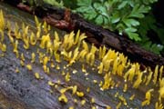 Steve Trudell |
102b Not erect, or if so, then expanding above into obconic shape, disc-shape, cup-shape, or flattened part (petal-like)
................................................................................103
103a Expanding above becoming obconic with the hymenium concave to convex, with or without stipe, or becoming cup-shaped
................................................................................104
103b Shaped otherwise (some of the sporocarps in a collection might be top-shaped, but sporocarps not uniformly obconic)
................................................................................108
104a Sporocarps growing on dead yellow-cedar foliage, twigs, and cones, near or under melting snow banks, (spores borne in asci)
................................................................................Gelatinodiscus flavidus
| ASCOCARP 1-5 mm across, up to 4 mm high, at first concave becoming convex when mature, even; bright to dingy yellow, drying dark olive; underside colored as upper surface; bald except for short yellow fuzz at base; stipe 2-5 mm long and about 1 mm wide. HABITAT on foliage, twigs, and cones of Chamaecyparis nootkatensis (yellow-cedar), consistently near or under melting snowbanks, April through August. MICROSTRUCTURES spores 26-34 x 9-11 um, oblong-ellipsoid with one end broader than the other, greenish yellow, with 2 droplets; asci 8-spored; paraphyses colorless, branched, curved at tips. | Gelatinodiscus flavidus A and O Ceska |
104b Sporocarps growing on other substrates
................................................................................105
105a Smooth to somewhat wrinkled on upper surface, undersurface whitish and finely pubescent to velvety, stipe short or absent; mostly on hardwoods; uncommon
................................................................................Femsjonia pezizaeformis
BASIDIOCARP 3-14 mm wide and about the same high, top-shaped to cup-shaped, basidiocarps compressed when clustered; smooth to somewhat wrinkled on upper surface, finely downy on undersurface; consistency gelatinous, soft; without stipe or with short stipe; light yolk-yellow (drying dull red-brown), margin white, outer surface whitish. HABITAT on hardwood, rarely conifer wood. MICROSTRUCTURES spores 22-27.5 x 7.5-11 um, curved-cylindrical, becoming tardily 3-many-septate at maturity, walls and septa thin; basidia forked; occasionally hyphidia; clamp connections present. REMARKS Also known as Ditiola peziziformis (Lev.) D.A. Reid. The appearance suggests an ascomycete, but microscopic differences obvious; most similar cups less gelatinous and more brittle. Heterotextus alpinus is smooth to pimply or ribbed on undersurface rather than velvety.
105b Exterior ribbed or at least roughened, but not whitish velvety
................................................................................106
106a Stipes of most basidiocarps more than half of the height of the basidiocarp; usually on hardwood; uncommon, any time of year but most often in fall, (exterior a palisade of thin-walled or thick-walled, septate, cylindrical, simple or branched hyphae in which the individual cells may become inflated)
................................................................................Guepiniopsis buccina
BASIDIOCARP up to 10 mm high, with head and stipe, closely gregarious or cespitose, both heads and stipes of adjacent sporocarps often partly fusing, head 3-9 mm across, cup-shaped or obliquely cup-shaped, externally longitudinally ribbed, stipe central and longitudinally ribbed; basidiocarp yellow to orange-yellow, drying bright orange, orange-red or rusty orange. HABITAT on hardwood or conifer wood. MICROSTRUCTURES spores 12-14.5 x 4-6 um, cylindrical to curved-cylindrical, becoming 3-septate at maturity, thin-walled with thin septa; basidia on inside of cup, shaped like tuning forks; exterior and stipe covered by a palisade of thin-walled or thick-walled, septate, cylindrical, simple or branched hyphae in which the individual cells may become inflated; clamp connections absent.
106b Stipes of most basidiocarps less than half the total height of basidiocarp; on conifer wood; common or uncommon, usually late winter and early spring, (exterior of thick-walled, obclavate, obovate, obpyriform or broadly cylindrical terminal cells), (Heterotextus)
................................................................................107
107a Some basidiocarps may be over 10 mm wide or over 8 mm tall; spores 14.5-18 um long and 3-septate
................................................................................Heterotextus alpinus
| BASIDIOCARP 3-10(25) mm wide and about the same high, with head and short central stipe, obconic with upper (spore-bearing) surface convex to concave and smooth or nearly so, externally ribbed, roughened with bumps; bright orange to pallid amber, drying dark orange-red. HABITAT scattered to gregarious, on coniferous wood in late winter or early spring, rarely in summer or fall. MICROSTRUCTURES spores 14.5-16.5(18) x 4.5-5.5 um, curved-cylindrical, becoming 3-4-septate, thin-walled with thin septa; conidia elongate, up to 4.5 x 1.5 um; basidia forked; clamp connections present. REMARKS Most cup fungi differ by being non-gelatinous, and microscopically most have asci. | Heterotextus alpinus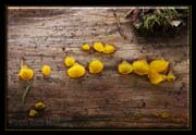 Rich Mably |
107b Up to 10 mm wide and up to 8 mm tall; spores 16-24 um long, becoming 5-7(9)-septate at maturity
................................................................................Heterotextus luteus
BASIDIOCARP up to 8 mm high, with head and stipe, head 4-10 mm in diameter, disc with head and short, stout central stipe, scattered to gregarious, obconic with upper (spore-bearing) surface convex to concave and smooth or nearly so, externally roughened; yellow to pallid yellow or lemon, drying more orange. HABITAT gregarious on conifer wood in late winter or early spring. MICROSTRUCTURES spores (16)17.5-22(24) x 4.5-5.5(6) um, curved-cylindrical, becoming 5-7(9)-septate, thin-walled with thin septa, basidia becoming forked; occasionally simple hyphidia; clamp connections present.
108a (103b) Upright, expanding away from substrate into flattened shape or funnel-shaped with indent or split on one side; solitary or in rows or clusters
................................................................................109
108b Not upright, shaped otherwise
................................................................................110
109a 2-8(18) cm high, higher than wide, tongue-shaped to somewhat funnel-shaped, usually indented or split on one side, with a head and stipe; pale to deep pink to reddish orange or salmon-colored; on rotting conifer wood or on ground under conifers, sometimes developing in lawns, or from buried wood near the edges of streams
................................................................................Guepinia helvelloides
(See 201a.)
109b 0.5-1.2(2.5) cm high, stipe slender, cylindrical at base, becoming flattened apically, there typically petal-shaped, spathulate or palmate, often deeply divided; yellow to orange, base of stipe often darkened; developing on hardwood or conifer wood, often sawn timber: cespitose, gregarious, or arranged in lines
................................................................................Dacryopinax spathularia
| BASIDIOCARP 0.5-1.2(2.5) cm high, with stipe and head 0.3-0.8(1.2) cm broad, stipe slender, cylindrical at base, becoming flattened apically, there typically petal-shaped, spathulate or palmate, often deeply divided, infrequently morel-like, consistency tough-gelatinous to cartilaginous, cespitose, gregarious, or arranged in lines; yellow to orange when fresh (drying yellow-brown to dull wine), base of stipe often darkened, brownish to gray or blackish; spore-bearing surface on one side or facing substrate, stipe and non-spore-bearing surface dull white, cream, or pallid tan; head and stipe tomentose. HABITAT on hardwood and conifer wood, often on fence posts or other sawn timber. MICROSTRUCTURES spores 7-11 x 3.5-4.5 um, slightly curved-cylindrical, becoming 1-septate at maturity; basidia forked, clamp connections absent, conidia up to 2.5 x 2.5 um. | Dacryopinax spathularia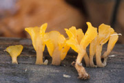 John Plischke |
110a (108b) Basidiocarps on dead plant material (including wood) or other nongilled fungi; without a distinct separate stipe: cushion-shaped, top-shaped, disc-shaped, cup-shaped, convoluted, folded, or with bladder-like lobes (includes Dacrymyces, Tremella)
................................................................................111
110b Basidiocarps growing from a distorted part (gall) of a living plant host, often horn-shaped (telial stages of Cronartium, Gymnosporangium), OR forming bumps on Gymnopus dryophilus (Syzygospora)
................................................................................134
111a Basidiocarps on recognizable fungal host
................................................................................112
111b Basidiocarps on wood or on a fungal host that is not recognizable
................................................................................115
It should be noted here that several Tremella species grow on lichens in the Pacific Northwest. None of these are more than 5mm across. Tremella cetrariicola grows on Cetraria and Tuckermannopsis, Tremella cladoniae on Cladonia, Tremella hypogymnia on Hypogymnia physodes, Tremella lethariae on Letharia vulpina, Tremella lichenicola on Mycoblastus fucatus, Tremella nephromatis on Nephroma, and Tremella papuana on Hypogymnia imshaugii and Hypogymnia pseudobitteriana . Some details are available on the MatchMaker CD (Gibson et al.), and full details in Diederich (1996, 2003).
112a Basidiocarps on hymenium of Acanthophysium lividocoeruleum basidiomes (the host grows in exposed habitats on conifer logs, often near streams)
................................................................................Tremella subencephala
BASIDIOCARP 0.5-3 mm wide, sometimes anastomosing and then up to 6 mm long and up to 2.5 mm high, cushion-shaped to nearly spherical, occasionally disc-shaped, typically aggregated, gelatinous but the larger basidiocarps often with a fleshy core, often appearing substipitate because of slight upward growth of host tissue, the basidiocarp often surrounded by a whitish ring of tufted hyphae on the host hymenium, the tufts consisting of mixed host/parasite hyphae; predominantly yellow. HABITAT Tremella subencephala is found on the surface of Acanthophysium lividocoeruleum, a crust species typical of rather dry habitats that grows on barkless wood. Basidiomes of the Acanthophysium are mostly blue-gray but some parts may be white. The Tremella is on the spore-bearing surface (the surface away from the wood), and is fairly conspicuous. Look for logs exposed to the sun, especially near streams. MICROSTRUCTURES spores 7.0-8.0 x 5.5-7.5 um, subglobose to broadly ellipsoid or ovoid, conidia abundant, 3.0-6.5 x 3.0-5.0 um, globose to ellipsoid; probasidia sparse, 10.5-14.0 x 10.0-11.0 um, borne on the same hyphae as conidia, subglobose to obovoid or ellipsoid, 4-celled, the walls often thickened at maturity; clamp connections present. REMARKS The tiny basidiocarps, known only on Acanthophysium lividocoeruleum, have been collected a few times in western Canada and Sweden. They are superficially similar to Tremella versicolor but the host is different and they are microscopically distinct: basidia of T. subencephala are smaller and they lack the basal stalk and swollen base that are common in T. versicolor, basidiospores have not been observed in T. versicolor, conidia are released singly in T. subencephala rather than in clusters as in T. versicolor, and individual conidia differ morphologically.112b Basidiocarps on hymenium of Aleurodiscus grantii basidiomes
................................................................................Tremella mycetophiloides
(See 503a.)
112c Basidiocarps on hymenium of, or adjacent to, the basidiocarps of Peniophora spp.
................................................................................113
112d Basidiocarps mostly adjacent to the host (Stereum hirsutum complex basidiomes) usually on hardwood, infrequently small basidiocarps develop on the host hymenium
................................................................................Tremella aurantia group
(See 120a.)
112e Basidiocarps mostly adjacent to the host (Stereum sanguinolentum basidiomes) on conifer wood, infrequently small basidiocarps develop on the host hymenium
................................................................................Tremella encephala
(See 613a.)
112f Basidiocarps arising from perithecial ostioles of Diaporthe and related species (1 basidiocarp per perithecium). The Tremella basidiocarp expands above and its source can only be seen in carefully prepared sections. (basidiospores globose)
................................................................................Tremella globispora
(See 507b.)
Note that the host basidiomes are very often unrecognizable, in which case the various species in this lead will key out below. Tremella versicolor, T. subencephala, and T. mycetophiloides always grow only on the host hymenium. T. mesenterica, T. mesenterella, T. aurantia, and T. encephala usually produce sporocarps on wood adjacent to fungal associates, but infrequently produce small basidiomes on the host sporocarp surfaces. There are many colonizers of recently dead branches or trees, and in the case of Tremella mesenterica, growth on the host may be no more than an accident caused by crowding. In Tremella aurantia, the Stereum is generally present only as a layer of hyphae within the T. aurantia lobes. In Tremella encephala, the Stereum forms a knob-like mass of hyphae surrounded by a thin gelatinous layer of the Tremella basidia and hyphae. Tremella foliacea basidiocarps contain hyphae of the Stereum or other host, but not in the proportions seen in T. encephala and T. aurantia. In the case of Tremella globispora, each basidiocarp arises from the perithecial ostiole of the host, expanding above and obscuring the presence of the host structure. Finally, some species of Tremella (e.g. T. subanomala) and Sirobasidium sporulate on or beside stromata of Diatrype bullata and related forms.
113a (112c) Basidiocarps 0.2-0.5 cm wide, cushion-shaped, hemispheric, discoid, infrequently anastomosing; usually developing from under bark; cream to pale yellow or dull orange when moist, the interior often brownish; uncommon
................................................................................Tremella versicolor
BASIDIOCARP 2-5 mm wide, cushion-shaped, predominantly hemispheric, the margins abrupt, sometimes disc-shaped, infrequently anastomosing to form extended, irregular masses, often concentrated on the edges of host basidiocarps or less often on the substrate adjacent to the host margin; cream to pale yellow or dull orange, the interior often brownish, drying yellowish tan to orange or brown. HABITAT fairly conspicuous on spore-bearing surfaces of Peniophora spp., which often occur near the bases of dead stems of Rubus and fallen Alnus branches, especially near the ocean, in situations where winter fog could be expected. MICROSTRUCTURES basidiospores not seen, conidia 4.0-6.5 x 4.0 x 5.5 um, globose to obovoid, in slide preparations conidia often released in clusters of 2-8, sometimes with a short length of their subtending hyphae; probasidia when present situated below the thick outer layer of conidia, (13)18-28(32) x 10-16(21) um, highly variable in shape, sometimes stalked, the stalk often tapering basally and then abruptly swollen near the base, 4-celled at maturity; clamp connections apparently present at all septa. REMARKS Tremella subencephala is superficially similar to Tremella versicolor q.v.
113b Basidiocarps reaching larger sizes, becoming lobed or brain-like; yellow, orange, flame-colored, red, maroon or brown
................................................................................114
114a Anastomosed masses up to 10 cm but often smaller; yellow to orange; spores 10-16 x 7-11 um: ovoid with length approximately one third greater than the width; species common on many domesticated and wild woody plant species
................................................................................Tremella mesenterica
(See 121a.)
114b Anastomosed masses up to 5 cm but often smaller; yellow, orange, flame-colored, red, maroon, buff, or brown; spores (11)12-15(16.5) x 10-12(14) um, globose or subglobose; species less common
................................................................................Tremella mesenterella
(See 121b.)
115a (111b) Most fresh basidiocarps yellow to orange (Tremella aurantia, T. mesenterella, T. mesenterica, Dacrymyces spp., Cytidia salicina)
................................................................................116
115b Fresh basidiocarps either with prominent brownish colors (but may be amber to yellowish brown) OR mostly ivory but may be yellow or yellow-tinged especially when young, or sometimes pinkish-tinged OR in small translucent droplets or cushion-shapes that may be yellowish but are predominantly whitish OR colorless to milky or pale pink or pale yellow growing on hymenium of Aleurodiscus grantii basidiocarps (these options including Exidia recisa, Tremella encephala, T. globispora, T. mycetophiloides)
................................................................................133
116a At least some basidiocarps (or their anastomosed masses) reaching 3 cm
................................................................................117
116b No basidiocarps or anastomosed masses reaching 3 cm
................................................................................122
Note: this is a helpful but not absolute distinction, since we are dealing with many sporocarps that have indeterminate growth that varies with rain, temperature, nutrients, etc.
117a Reddish orange to wine-red basidiocarps that start as resupinate discs up to 1 cm across, thin-fleshed, loosening along margins to become shallow cups, often confluent for several decimeters, on hardwoods especially Salix (willow)
................................................................................Cytidia salicina
(For description, see 207a.)
117b Yellower orange, or not forming cups, or growing on conifer wood (any of these)
................................................................................118
118a Basidiocarps on conifer wood (with or without fungus host), or indeterminate wood
................................................................................119
118b Basidiocarps on hardwood (with or without visible fungus host)
................................................................................120
119a With white point of attachment; common, on conifer wood
................................................................................Dacrymyces chrysospermus
(a synonym is Dacrymyces palmatus)
| BASIDIOCARP variable in shape, cushion-shaped, fan-shaped, or stoutly stipitate with spatula-shaped, cup-shaped, plate-shaped, or convoluted head, sometimes as a cluster of 2-3 almost coralloid lobes; often coalescing to form erect, brain-shaped or complicated sessile or stoutly stipitate masses up to 6 cm across; attached by tough, white, rooting base, stipe and base of head often with hairs; bright yellow orange to orange when fresh, drying orange-red to orange-brown. HABITAT normally on conifer wood, may possibly occur on hardwood. MICROSTRUCTURES Spores are 16.5-23(26.5) x 5-7.5 um, curved-cylindrical, relatively thin-walled with slightly thickened and distinct septa, becoming 7-septate by maturity. Colorless ovoid conidia may be present. Basidia are shaped like tuning forks. Occasionally there are simple hyphidia. Hyphae are septate without clamp connections. This species often has an internal Tremella parasite (possibly more than one), especially noted when there appears to be deliquescence at the edge of the basidiocarp. Not only Tremellaceous parasites but some of the simple-pored forms with auricularioid basidia (formerly Platygloeales) parasites attack species in Dacrymycetaceae. REMARKS Dacrymyces chrysospermus is known in most field guides as Dacrymyces palmatus and is one of the most common jelly fungi in conifer forests. Tremella mesenterica lacks white basal attachment and generally grows on deciduous wood (rarely on pines). T. mesenterica generally develops on trunks and branches that still have bark, mostly arising from a large, tough, gelatinous primordium beneath the bark, and also differs microscopically from D. chrysospermus. The other Dacrymyces species are smaller and simpler in form, less common, and differ microscopically. Dacrymyces capitatus is often convoluted but is ivory to yellow, usually smaller, usually grows on hardwoods, and has different spores. | Dacrymyces chrysospermus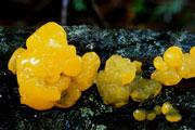 Norman Evans |
119b Sessile, without white point of attachment; mostly on hardwoods
................................................................................120
120a (118b, 119b, 123b) Fleshy whitish layer plainly visible on section through basidiocarp; (spores 7.5-10 x 6-8 um, unclamped Stereum hyphae present)
................................................................................Tremella aurantia group
| BASIDIOCARP usually 2-5 cm across and 1-4 cm high, (but sometimes on red alder approaching 20 cm in diameter), densely lobed, brain-like, folded (folds often becoming leaf-like); bright orange or orange-yellow, drying ochraceous to red-brown; whitish fleshy internal layer (representing Stereum host) often visible on section through basidiocarp (slice lobes vertically lengthwise) and may form cottony masses; lobes may be hollow. HABITAT This group is usually reported on hardwood, less often conifer wood. The species is a parasite of Stereum basidiomes. Basidiocarps of the Tremella are only infrequently seen on the hymenium of the Stereum, generally simply occurring beside the host and having a zone of host hyphae internally. In BC small basidiocarps are sometimes seen on recognizable Stereum (on oak). Although basidiocarps of this group can be found on fallen trunks and branches, the easiest place to find them is on the dead lower branches of suitable substrate trees, accompanied by Stereum species. MICROSTRUCTURES spores 7-10 x 6-10 um, ellipsoid to subglobose, basidia 2-4-spored, most with diagonal septa; with clamp connections, intermixed with Stereum host hyphae which lack clamp connections; haustorial cells present and conspicuous, mostly spherical, 2-3 um wide, giving rise to one or more thin filaments. REMARKS The are two members of this group in British Columbia that superficially resemble T. aurantia Schw.: Fr. Their identity is uncertain at this time. They differ from each other in DNA, some morphological characters, and substrate, and are not conspecific. On Vancouver Island, one develops on Quercus garryana (Garry oak), especially on dead, attached lower branches. Lobes are often inflated and hollow, the hollow area lightly stuffed with white hyphae (presumably of the associated Stereum hirsutum group). Sometimes small basidiomes are produced on Stereum. That Tremella taxon appears to be close to T. aurantia in the sense of Schweinitz. The other taxon occurs on standing or fallen Alnus rubra (alder). It is close to the true Tremella aurantia in form, but does not appear to be the same. These two taxa belong to a group including T. aurantia, T. encephala, T. australiensis, T. tremelloides ("Neotremella" Lowy), T. aurantilutea Bandoni & Zang, and other species including some sporocarpless endoparasitic taxa listed by C-J. Chen, 1998. | Tremella aurantia.jpg) Michael Wood (MykoWeb) |
120b Whitish layer not plainly visible on section through basidiocarp; (spores 10-16 x 7-12 um)
................................................................................121
121a Anastomosed masses up to 10 cm but often smaller; yellow to orange; spores 10-16 x 7-11 um: ovoid with length approximately one third greater than the width; species common
................................................................................Tremella mesenterica
| BASIDIOCARP 1-10 cm, brain-like or with irregular clustered folds consisting of one to several distorted lobes, these typically arising from a large, tough, gelatinous, subcortical primordium with rounded edges, and breaking through the bark, sometimes in several places, the lobes 0.5-0.8 cm x 0.3-0.7 cm in individual basidiocarps, anastomosing up to 10 cm x 5 cm; no stipe; golden yellow, yellow-orange, orange, pale yellow, more rarely whitish or almost colorless; drying red to orange or brown. HABITAT on hardwood or uncommonly conifer wood (e.g. pine), associated with Peniophora spp., mostly on recently dead branches that still have bark, infrequently on barkless patches of such branches. Rarely small basidiomes are seen growing right on the associated Peniophora, but there may be no obvious connection with living Peniophora basidiocarps, especially as many other fungal species share these habitats. The maximum numbers of basidiocarps are seen in young alder thickets, in which deaths of some of the crowded trees, and many branches, is occurring. MICROSTRUCTURES Basidiospores are (9.5)10.5-16 x (7.5)8.5-10.5(11.5) um, (Wong), 7-18 x 6-14 um (Arora), broadly ellipsoid, obovate, or subglobose, basidia longitudinally to obliquely septate, 4-spored. The hymenium sometimes consists only of conidia, especially in winter or early spring, but it will later contain both conidia and basidia, or finally only basidia, the latter forming first beneath the conidial layer if that is present. Conidia are 3-5 x 2-3 um, ellipsoid to subglobose; conidial state is often borne in cool periods in fall, winter, and spring; Hyphidia are absent, or sometimes in early stages of development with some hyphidia (but not thick-walled as in T. mesenterella). Clamp connections are present throughout. REMARKS Tremella mesenterica is common in the Pacific Northwest and is found year-round in wet weather on many cultivated and wild species of woody plants but is rare on conifer wood. Dacrymyces chrysospermus has a white point of attachment and grows primarily on coniferous wood. | Tremella mesenterica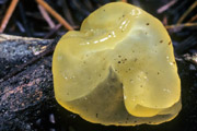 Michael Beug |
121b Anastomosed masses up to 5 cm but often smaller; yellow, orange, flame-colored, red, maroon, buff, or brown; spores (11)12-15(16.5) x 10-12(14) um, globose or subglobose; species less common
................................................................................Tremella mesenterella
| BASIDIOCARP up to 5 cm long, 1.5 cm wide, and 1.3 cm high, often beginning as a lobe protruding through ruptured bark (arising from a primordium under the bark), developing leaf-like lobes or becoming brain-like or top-shaped; dull yellow, orange, flame-colored, red, maroon, brown, or buff, sometimes minutely punctate with darker brown spots; drying mostly dark red, brown, or almost black; basidiocarp often bearing a thick layer of conidiogenous cells and conidia and then opaque rather than translucent as with basidiocarps. HABITAT most frequently on dead attached lower branches of Cornus (dogwood) and Salix (willow) spp., often in association with basidiocarps of Peniophora spp. (infrequently producing small basidiocarps on the Peniophora hymenium), from late fall to early spring, or in summer. MICROSTRUCTURES basidiospores (11)12-15(16.5) x 10-12(14) um, mostly bulliform, some subglobose, the apiculus midway on the flattened surface in the bulliform ones; basidia 4-celled; well-developed thick-walled sterile hyphidia; clamp connections abundant. REMARKS Specimens from different substrates differ in some respects: for example those on Salix from BC and on Cornus and Hamamelis (witch-hazel) from NC are commonly brownish (brown to maroon or red) rather than pale dull yellow to light brown as in specimens from Cornus in BC. They are probably distinct species. On Salix basidiomes may be found side by side with Tremella mesenterica and with associated Peniophora basidiomes. | Tremella mesenterella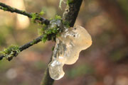 A and O Ceska |
122a (116b) Gregarious on sodden coniferous wood, often on sawn timber, on the undersurface of floating wood or more often at or near the waterline on partially exposed wood; typically less than 1 mm in diameter, sometimes confluent and reaching 3-4 mm across; (spores 8.5-11(12) x 3.5-4.5 um, curved-cylindrical, arthoconidia 4-6(6.5) x 2.5-3.5 um produced by fragmentation of hymenial hyphae)
................................................................................Dacrymyces aquaticus
BASIDIOCARP typically less than 1 mm in diameter, gregarious, sometimes confluent and reaching 3-4 mm across, about 0.5 mm high, cushion-shaped, smooth or slightly roughened, attached weakly by a point; pallid yellowish becoming yellow or orange. HABITAT on sodden coniferous wood, often on sawn timber, common on the undersurface of floating wood, but more often at or near the waterline on partially exposed wood. MICROSTRUCTURES spores 8.5-11(12) x 3.5-4.5 um, curved-cylindrical, aseptate when shed and remaining so at germination or, more often, becoming 1-septate, rarely 2-septate; basidia forked, sparse; no conidia are formed on spores or germ tubes; arthoconidia 4-6(6.5) x 2.5-3.5 um, ellipsoid, 1-celled, produced by fragmentation of hymenial hyphae; clamp connections absent.
122b Not showing above characters
................................................................................123
123a Basidium aseptate, consisting of 1 cylindrical to clavate cell from the apex of which arise 2-4 large, conspicuous epibasidia (not separated from the cell by a septum) each bearing a single spore (Dacrymyces)
................................................................................124
123b Basidium divided by longitudinal or occasionally irregular septa into (2, 3) 4 cells, each of which bears a single spore (Tremella)
................................................................................120
Note that Dacrymyces species may occasionally be parasitized by Tremellaceous fungi or species of Platygloeales sensu lato, so that some foreign basidia may be present.
124a Spores predominantly 16-26 um long or longer, cylindrical and may be curved, becoming 7-septate by maturity
................................................................................125
124b Spores shorter, or less septa at maturity
................................................................................127
125a Basidiocarps extremely variable in shape, cushion-shaped, fan-shaped, stipitate with a head that is spatula-shaped, cup-shaped, convoluted, roughly coralloid, or plate-like; clamp connections absent or obscure
................................................................................Dacrymyces chrysospermus
(See 119a.)
125b Basidiocarps cushion-shaped to cup-shaped; clamp connections present
................................................................................126
126a Spores 24-28 (32) x 7.5-10(11) um, becoming 7-septate, consistently becoming thick-walled with thickened septa, often with 1-3(5) longitudinal septa
................................................................................Dacrymyces chrysocomus
BASIDIOCARP 1-4 mm across, up to 2.5 mm high, at first cushion-shaped, becoming centrally depressed, finally cup-shaped, externally with hairs, with little or no stipe, gregarious; surface yellow to orange-yellow, drying reddish brown. HABITAT on conifer wood. MICROSTRUCTURES spores 24-28(32) x 7.5-10(11) um, fusiform but occasionally subglobose to broadly ellipsoid (then 14-20 x 10-16 um), becoming thick-walled with thick septa, transversely 7-septate with 1-3(5) longitudinal septa, (McNabb), 17-23.5 x 6.5-8.5 um, almost cylindrical, (Raitviir), 16.0-24.0 x 7.75-8.75 um, varying from ellipsoid to slightly allantoid, becoming transversely 3-7(8)-septate and at maturity developing also a number of longitudinal septa and so appearing strikingly muriform, (Reid); basidia forked; occasionally simple hyphidia; clamp connections present. REMARKS The differentiation from Dacrymyces variisporus follows McNabb (1973): the differentiation becomes more difficult if the spore measurements of Raitviir or Reid are used for Dacrymyces chrysocomus.
126b Spores (12) 16-26 (30) x 6-9.5 um, becoming 3-7-septate, thin-walled or thick-walled with irregularly thickened septa, occasionally with 1-2 longitudinal septa
................................................................................Dacrymyces variisporus
BASIDIOCARP 0.5-5 mm across, up to 2 mm high, at first pustulate, becoming top-shaped or cushion-shaped and centrally depressed, smooth to slightly wrinkled-pleated, gregarious, occasionally coalescing to form masses to 15 mm in extent, broadly attached by center of undersurface; pallid orange to orange-brown or yellow or fading to nearly colorless, drying amber to orange, orange-red, or dark brown. HABITAT on conifer wood and hardwood. MICROSTRUCTURES spores (12)16-26(30.5) x 6-9.5 um, curved-cylindrical, occasionally fusiform, often thick-walled with irregularly thickened septa, becoming (3)7-septate at maturity, occasionally with 1-2 longitudinal septa, colorless ovoid conidia may be present; basidia forked; simple hyphidia present often with 1-3 clamp connections throughout their length; internal hyphae with clamp connections present.
127a (124b) Spores in chains (arthrospores), 8-16 x 2.5-5.5 um, these sporocarps opaque, slimy, bright orange or reddish orange when fresh, drying dark red-orange, hemispherical, irregular or tuberculate, slimy rather than gelatinous, typically but not always accompanied by gelatinous basidiocarps of same species that are gelatinous, pale yellow to orange-yellow when fresh, drying yellow to yellow brown, pustulate, disc-shaped, convoluted or cerebriform, or the arthrospores occurring in the basidiocarp
................................................................................Dacrymyces stillatus
See 131a for description.)
127b Spores subglobose to broadly ovate, or curved cylindrical
................................................................................128
128a Spores subglobose to broadly ovate, initially becoming 4-celled and resembling Tremellaceous basidia
................................................................................Dacrymyces ovisporus
| BASIDIOCARP 2-5 mm in diameter, to 4 mm high, pustulate, convoluted with gyrose lobes and folds, orange when fresh, drying dark amber to dark brown. HABITAT on conifer wood. MICROSTRUCTURES spores (9.5)13-15 x 8-12 um, subglobose to broadly ovoid, thin-walled with thin septa, spore initially becomes divided by a single transverse, longitudinal, or oblique septum, then two secondary septa form at right angles to the first, and finally some thin irregularly placed septa; colorless, globose conidia; basidia forked; simple hyphidia present; clamp connections present. REMARKS Spores of D. chrysocomus are occasionally similar in shape and have septa in more than one plane, but the majority of spores are broadly and bluntly fusiform. Reid (1973) says spores of D. ovisporus are 16-17 x 9.75-11.75 um, mostly ovoid but sometimes ellipsoid. | Dacrymyces ovisporus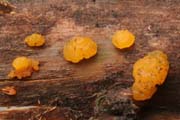 A and O Ceska |
128b Spores curved cylindrical
................................................................................129
129a 0.8-4 mm in diameter, about the same in height, cushion-shaped, top-shaped or cup-shaped
................................................................................Dacrymyces minutus
BASIDIOCARP 0.8-3 mm in diameter, about the same in height, cushion-shaped, top-shaped, or shallowly cup-shaped, occasionally irregular, substipitate or stipitate, stipe rarely to 3 mm long, gregarious; dull orange to bright orange-yellow, drying reddish orange to orange-brown. HABITAT gregarious on conifer wood. MICROSTRUCTURES spores 13-18(19.5) x 4.5-6(7) um, curved-cylindrical, thin-walled with thin septa, becoming 3-septate at maturity, colorless conidia may be present; basidia forked; occasionally simple hyphidia; clamp connections present; cortex covered with thick-walled, simple or branched, clamped hairs.
129b Larger or more variable in shape
................................................................................130
130a Clamp connections present
................................................................................Dacrymyces tortus
BASIDIOCARP 0.5-2 mm across, up to 2 mm high, pustulate, becoming cushion-shaped and centrally depressed, sometimes convoluted, gregarious, typically discrete, occasionally coalescing to form convoluted areas up to 20 mm in extent, attached to substrate by central point; amber or dingy yellow, often with greenish tints, sometimes appearing pallid to almost colorless, drying dark amber to brown or dull black. HABITAT on coniferous wood. MICROSTRUCTURES spores (8)10-14(15) x 3.5-4.5(5) um, slightly curved-cylindrical, thin-walled with thin septa, becoming tardily 1-3-septate at maturity; basidia forked; simple hyphidia present with 1-3 clamp connections throughout their length; clamp connections present on internal hyphae. REMARKS Note that G.W. Martins concept of Arrhytidia involuta (Schwein.) Coker, considered by McNabb (1973) and Ginns & Lefebvre (1993) a synonym of Dacrymyces capitatus Schwein., would key out here, because unlike McNabb, he found conspicuous clamp connections. Dacrymces stillatus could also key out here if there are clamp connections, as claimed by some authors, but D. stillatus has hyphidia that are lacking or scarcely distinguishable whereas D. tortus has septate hyphidia exceeding the basidia and with clamp connections . Dacrymyces minor usually grows on hardwood, and it does not have the conspicuous clamp connections. Note that Guepiniopsis torta Pat. is a synonym of Guepiniopsis buccina (Pers.:Fr.) Kennedy, not of Dacrymyces torta.
130b Clamp connections absent
................................................................................131
131a Spores distinctly thick-walled, septa thickened
................................................................................Dacrymyces stillatus
| BASIDIOCARP 1-4(15) mm across, up to 3.5 mm high, cushion-shaped, knob-shaped, lens-shaped, cup-shaped, or plate-shaped, with a stipe-like attachment in center, yellow to orange-yellow, drying yellow-brown to dark orange or red-brown; smooth to undulating-wrinkled, sometimes also slightly brain-like, sometimes coalescing to form masses up to 20 mm in extent; arthrospore-bearing fructifications hemispheric, irregular, or tuberculate, bright orange or reddish orange, opaque, drying dark red-orange. HABITAT on conifer wood, less often hardwood, fairly common on dead culms of Sambucus pubens (red elderberry). MICROSTRUCTURES basidiospores (9)10.5-16(17.5) x 3.5-6 um, curved-cylindrical, occasionally ovate to subpyriform (nearly pear-shaped), becoming thick-walled with thick septa, 3-(4)-septate at maturity; colorless, globose to subglobose conidia up to 4 um x 2 um may be present; basidia forked; occasionally simple hyphidia but not conspicuous; clamp connections not seen in any specimens by McNabb (1973) and Kennedy (1958b), and only one clamp in many collections by R. Bandoni (pers. comm.), but presence recorded by other authors; arthrospores often present in separate sporocarps or occasionally mixed with basidia: outer layers of arthrospore stage composed of chains of arthrospores, (1)-2-celled, 8-16 x 2.5-5.5 um. REMARKS Dacrymyces tortus has clamped hyphidia and narrower spores. Dacrymyces capitata has short substantial stipe and somewhat smaller and thin-walled spores. Dacrymyces minor is usually on hardwoods, and has spores up to 4-cellular with thin septa. | Dacrymyces stillatus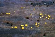 Steve Trudell |
131b Spores thin-walled, septa thin
................................................................................132
132a Basidiocarps sessile, pustulate or cushion-shaped, 0.5-2mm in extent
................................................................................Dacrymyces minor
| BASIDIOCARP at first pustulate, becoming cushion-shaped to disc-shaped, 0.5-3 mm wide, often in crowded groups which may coalesce somewhat and reach 10 mm in extent, smooth or centrally depressed and slightly convoluted, rarely brain-like, attached to wood by a central point; dingy orange or yellow, often with olivaceous tints when young, drying amber. HABITAT gregarious, on hardwood, less often conifer wood. MICROSTRUCTURES spores 8-14(15.5) x 3.5-5(6) um, curved-cylindrical, becoming 1-3-septate at maturity, typically thin-walled with thin septa, occasionally walls and septa slightly thickened; colorless spherical to ovate conidia may be present up to 2.5 um diameter; basidia forked; occasionally simple hyphidia; hyphae septate without clamp connections. REMARKS Dacrymyces minutus is even smaller and is sometimes irregular in shape, but its spores are larger. Dacrymyces stillatus is larger, has spores that are thick-walled with thick septa, and possesses an arthrospore state. | Dacrymyces minor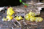 Michael Beug |
132b132b Basidiocarps often short-stipitate, attached by whitish rooting base, variable in shape and 0.5-20 mm in extent
................................................................................Dacrymyces capitatus
| BASIDIOCARP 0.5-15 mm across, up to 5 mm high, cushion-shaped to plate-shaped or concave, smooth, gyrose, or convoluted, usually with short or rudimentary, somewhat rooting stipe, gradually merging into upper part of basidiocarp, stipe and base of head covered with hairs, basidiocarps often coalescing to form masses 20 mm across; pale yellow to orange-yellow or brownish orange, sometimes white-pruinose, drying brown to brownish red. HABITAT dead wood with and without bark. MICROSTRUCTURES spores 11-17 x 3.5-6(7) um, curved-cylindrical, 3-septate at maturity, typically thin-walled, occasionally walls and septa slightly thickened; basidia forked; occasionally simple hyphidia; clamp connections absent. REMARKS Dacrymyces stillatus lacks a substantial stipe and has larger, more thick-walled spores. | Dacrymyces capitatus A and O Ceska |
133a (115b) Color varies from ivory, to pale tan, yellowish brown, yellowish, white, or colorless, sometimes with a pinkish tinge from associated Stereum
................................................................................Tremella encephala
(See 613a.)
133b In small translucent droplets or cushion-shapes that may be yellowish but are predominantly whitish; uninfected perithecia of the Valsaceous hosts typically present among basidiocarps, (spores globose)
................................................................................Tremella globispora
(See 507b.)
133c Colorless to milky or pale pink or pale yellow on spore-bearing surface of Aleurodiscus grantii
................................................................................Tremella mycetophiloides
(See 503a.)
133d Basidiocarps amber, yellowish, brown, cinnamon, pale liver brown, dark red-brown; mostly obconic when separate, but crumpled when clustered with little anastomosis, may be plate-shaped, conchate, wavy, brain-like, lobed; on hardwoods, often Salix
................................................................................Exidia recisa
(See 609a.)
134a (110b) Forming bumps on Gymnopus dryophilus
................................................................................Syzygospora effibulata
(See 604a.)
134b Basidiocarps growing from a distorted part of a living plant host, often horn-shaped (telial stages of Cronartium, Gymnosporangium)
................................................................................135
135a On conifers; teliospores are mostly 2-celled, borne singly on gelatinizing pedicels
................................................................................Gymnosporangium spp.
135b On dicotyledonous angiosperms; teliospores are 1-celled, borne in chains
................................................................................Cronartium spp.
PURPLE OR PINK
201a 2-8(18) cm high, higher than wide, tongue-shaped to somewhat funnel-shaped, usually indented or split on one side, with a head and stipe; pale to deep pink to reddish orange or salmon-colored; on conifer wood or on ground under conifers
................................................................................Guepinia helvelloides
| BASIDIOCARP 2-8(18) cm high, higher than wide, tongue-shaped to somewhat funnel-shaped, usually indented or split down one side, flabby-rubbery, with a head and off-center or lateral stipe, single to cespitose, inner surface smooth and dull, sometimes whitish-pruinose, outer surface smooth, often wrinkled-veined in age, hymenium on upper part of outer surface; pale to deep pink to reddish orange or salmon-colored. HABITAT on rotting wood or on the ground under conifers (perhaps on buried wood), sometimes developing in lawns, or from buried wood in edges of streams; late summer and fall, rarely spring. MICROSTRUCTURES spores 9-12 x 4-6.5 um, oblong to ellipsoid; basidia longitudinally septate, 2-4-spored; clamp connections present. | Guepinia helvelloides A and O Ceska |
201b Smaller or differently shaped, or lacking stipe, or on hardwood
................................................................................202
202a Top-shaped to disc-shaped or cup-shaped ascocarp that is purple to pink, with stipe lacking or short; tightly clustered growth on wood; common; (ascospores 10-19 um long that are nonseptate to 1-septate)
................................................................................Ascocoryne sarcoides
| ASCOCARP 0.5-1.0 cm across, cushion-shaped, top-shaped, cup-shaped, or disc-shaped, often irregular and lobed, clustering to 15 cm across, sessile or with very short stipe, surface smooth to wrinkled, exterior smooth; flesh-pink to violet-pink or reddish purple. HABITAT stumps and fallen logs and branches. MICROSTRUCTURES spores 10-19 x 3-5 um, ellipsoid, with single septum when ripe, two droplets; asci 8-spored; paraphyses often abruptly swollen at tip; ascocarps often accompanied by similarly colored conidial state. REMARKS There are usually accompanying conidiomata (Coryne dubia), also gelatinous in consistency, that are that are of variable form, often pustulate to irregularly clavate and a similar amethyst or pale lavender color, and may be opaque because of surface conidia. Ascocoryne cylichnium is very similar to A. sarcoides but often has larger ascocarps and is distinguished by larger spores, 18-30 x 4-6 um, which become multiseptate. | Ascocoryne sarcoides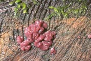 Sharon Godkin Ascocoryne cylichnium 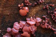 Rich Mably |
202b Saucer-shaped to convoluted brain-like ascocarp, purple brown (paler or pinkish when young), rare, (ascospores 6-10 x 3.5-5 microns)
................................................................................Ascotremella faginea
See 605b
202c Other shapes or colors, if cup-shaped or disc-shaped then either wine-red (to reddish orange) and thin-fleshed, or growing on shredded inner bark of Populus or growing on Prunus spp.
................................................................................203
203a Basidiocarps variously formed, spread out flat and irregularly rounded, brain-like, mesentery-like, or weakly cup-shaped; ocher-pink, grayish ocher, to salmon-colored; basidiocarps initially petal-like, one or more arising from edges of conidial sporocarps that are minute, bright pink to orange, and shaped like complex cups; in Europe on Prunus spp., in British Columbia growing on shredded inner bark of Populus trichocarpa
................................................................................Craterocolla cerasi
BASIDIOCARP 1-5 cm across, variously formed: petal-like, weakly cup-shaped, spread out flat and irregularly rounded, lobed, foliaceous, brain-like, or mesentery-like; sometimes short-stipitate, upper surface smooth, consistency gelatinous-glutinous; ocher-pink or gray-ocherish or salmon-colored. Conidiomata, usually appearing as aggregates before the basidial stage, are minute, bright pink to orange, complex cups: stalked, top-shaped, urceolate, with a membranous, stellate closure, colored as basidial form but not gelatinous, small but clustered and conspicuous. The basiocarps often develop as paler, spathulate or rounded lobes from one edge of a conidioma, resembling an Exidia or Tremella, petal-like in form at first but soon obscuring their origin and becoming clustered. HABITAT near Ladner in southern British Columbia found on bark of Populus trichocarpa, especially well-weathered (shredded) inner bark that has fallen away from dead trunks and has become trapped on shrubs below in cold winters (especially those with snow). It is also present on the inner layer of bark on the ground and it has been observed on the pith of the sawn end of a log. It is not certain that this is the same species as Craterocolla cerasi (Schumach.) Bref. which occurs in some areas of the world on dead trunks and branches of Prunus spp., occasionally other hardwoods. MICROSTRUCTURES spores 8-11 x 3.5-4.5 um, curved-cylindrical, conidia 6-8 x 2.5-3.5 um; basidia longitudinally septate, 4-spored; cystidia not seen; no clamp connections. REMARKS Craterocolla cerasi differs from Tremella by the separate occurrence with C. cerasi of conidiomata which usually appear before the basidium stage.
203b Other shapes or colors, growing on hardwood or conifer wood
................................................................................204
204a Hemispheric cushion-shaped or brain-like, with whitish fleshy core surrounded by a gelatinous layer that varies from pale pinkish brown, yellowish brown, or pale tan, to pinkish, yellowish, white or colorless (the pinkish color probably from pigment produced by host); on dead conifer wood or less often hardwood, associated with Stereum sanguinolentum
................................................................................Tremella encephala
(See 613a.)
204b Other shapes or colors; most species on hardwood (T. foliacea also on conifer wood)
................................................................................205
205a 2.5-20 cm wide or more, typically a roundish mass of wavy or leaf-like folds, lobes, and convolutions; reddish cinnamon, brown, vinaceous brown, purplish brown, or blackish brown; surface smooth, sporocarps sometimes split irregularly above
................................................................................Tremella foliacea
(See 614a.)
205b Differently shaped or colored
................................................................................206
206a Thin-fleshed, hymenium wine-red to dull purple (to reddish orange) basidiocarps that start as resupinate discs up to 3cm across and become shallow cups, often confluent for several decimeters, on hardwoods especially Salix (willow) and bark of Betula (birch)
................................................................................207
206b Thick-fleshed, or differently colored
................................................................................208
207a Usually on Salix (willow), hymenium wine-red (to reddish orange), (spores 12.5-17.5 x 4-5 um, hyphidia present in hymenium but no cystidia)
................................................................................Cytidia salicina
BASIDIOCARP at first disc-shaped pressed against wood, loosening along margin to form shallow cups, irregularly rounded to oblong, 0.5-1.0cm in size, with point-like attachment to the wood, later coalescing to form bands up to several decimeters long, up to 0.5-0.8 mm thick, elastic, leathery to gelatinous, tough, when dry shriveled, crustose, and hard; bright orange or orange-red to dark red, violaceous red or wine-red, drying dull wine-red; when moist smooth, pruinose and dull, slightly warty; abhymenial side covered by crystals and therefore whitish, farinose or finely floccose. HABITAT on attached dead or damaged branches with bark, as well as standing trunks, of Salix (willow) and other hardwoods, maturing in fall. MICROSTRUCTURES spores 12.5-17.5 x 4-5 microns, cylindrical, slightly allantoid, smooth, inamyloid, colorless, with many small droplets; basidia 4-spored, large, protruding from dense layer of dendrohyphidia that are 2.5-3 microns wide, very densely interwoven and imbedded in a gelatinous substance, strongly branched, brownish, +/- thick-walled; no cystidia; septa with clamps.
207b On a variety of hardwoods, hymenium pink to reddish brown, (spores 10-15 x 4-5 um, hyphidia and cystidia in hymenium)
................................................................................Cytidia patelliformis
BASIDIOCARP resupinate to open cup-shaped, up to 2.0 cm long and 1 cm wide, some confluent, hymenial surface pink to dark reddish-brown, pulverulent (powdery) to setulose under hand lens, surface next to wood yellow-buff to dark reddish-brown, with matted white hairs when well-developed. HABITAT on fallen branches of various hardwoods, August to April. MICROSTRUCTURES spores 10-15 x 4-5 microns, slightly curved, smooth, colorless; basidia 4-spored, often projecting 10-11 microns above hymenial surface; dendrohyphidia intermingled with basidia, 2-3 microns wide; cystidia projecting up to 60 microns beyond hymenial surface, 7-8 microns wide, cylindric or tapering slightly, a few with swollen apices; hyphae of context with walls somewhat thickened and strongly gelatinized, with numerous and prominent clamp-connections.
207c Known only from type specimen in Idaho on bark of Betula (birch), hymenium dull purple, spores 6-7 x 2-3 um, no sterile elements in hymenium
................................................................................Cytidia lanata
BASIDIOCARP 1-3cm across, attached at and near a central point, free over remainder of surface next to wood, free surface covered with tan to brownish woolly tomentum, more or less zonate; hymenium dull purple, smooth to somewhat wrinkled. HABITAT type scattered on bark of branch of Betula. MICROSTRUCTURES spores 6-7 x 2-3 microns, ovate to oblong-elliptic, smooth, colorless; no sterile elements seen in context or hymenium; hymenium in 2 or 3 layers, hyphae of context gelatinous and without clamp connections at the infrequent septa.
208a (206b) Up to 20 mm across, somewhat resembling a mulberry or blackberry; dark reddish brown to almost black, discoloring the surrounding barked hardwood it grows on
................................................................................Tremella moriformis
BASIDIOCARP 0.2-1.8 cm across, 0.1-0.8 cm high, starting as gregarious erumpent pustules which become confluent, then moriform (like a mulberry fruit); dark reddish brown or dark purplish brown or dark violaceous to almost black, drying black; in section, the interior reddish to purplish, the spore-bearing surface almost black, often discoloring the substrate with a purple or rose pigment; there is often a hard blackish core, possibly produced by a pyrenomycetous host. HABITAT on barked dead branches, recorded on hardwood. MICROSTRUCTURES spores 7.5-11 x 7-10 um, globose or more often broader than long, with apiculus at right angles to the long axis; basidia longitudinally to obliquely cruciate-septate, 4-spored; clamp connections present; globose to obovate or pyriform conidia, 3-8.4 x 2.3-6.1 um.
208b 5-25 mm across, with central point of attachment, remaining discrete disc-shaped to top-shaped, or anastomosing to form convoluted masses up to 80 mm long; at first grayish lilac, becoming brownish gray to dark brown with olive to violaceous tints, when slightly dry appearing blackish brown; surface usually roughened by conic papillae
................................................................................Exidia zelleri
BASIDIOCARP at first pustulate, expanding, 5-25 mm across, with a central to lateral point of attachment, remaining discrete disc-shaped to top-shaped, or anasotomosing to form convoluted masses up to 80 mm long appearing broadly attached by closely spaced points of attachment; outer surface flat to ridged to convoluted or lobed, marked by sparse to numerous conic papillae, undersurface free or lightly appressed, usually with small elevations or with vein-like to gill-like elevations, margin occasionally with papillae; at first grayish lilac, becoming brownish gray to dark brown with olive to violaceous tints, when slightly dry appearing blackish brown. HABITAT growing on hardwood, for example growing in early spring on vine maple wood on the ground. MICROSTRUCTURES spores (14.5)16-19(21) x 5-6 um, curved-cylindrical, basidia longitudinally to obliquely septate into 4 cells; conspicuous clamp connections present. REMARKS Exidia repanda is similar in form but different in color (brownish translucent, pinkish brown, cinnamon brown, gray brown, olivaceous brown, or yellowish brown). Exidia glandulosa lacks violaceous tints in the fresh basidiocarps, and has smaller spores. An unpublished Exidia species, described by Klett (1962) from willow in Washington, British Columbia, and Alaska, lacks violaceous tones, has a surface that is smooth to finely asperulate instead of being marked by conic papillae, and has generally larger spores, (17)18.5-22.5(25) x 5-6 um.
GREEN
301a Starting as pustules, maturing as single or occasionally multiple lobes, surface becoming smooth or convoluted, coalescing to form irregular areas up to 10 cm across; almost colorless, dingy translucent, dingy white, or pale yellow-brown, then when fully developed with a greenish or olivaceous tint, drying to a brown film; margin abrupt; (spores 9.3-13(15.4) x 5-8.7 um, gloeocystidia present and becoming yellowish granular but not projecting)
................................................................................Sebacina lactescens
(often known as Ductifera sucina)
BASIDIOCARP originating as colorless (hyaline) pustules up to 2 mm across, maturing as single or occasionally multiple lobes, becoming smooth or convoluted to brain-like, coalescing to form irregular areas up to 10 cm long, thickness 0.3-1.2 cm, consistency waxy-gelatinous; colorless almost like a thin piece of ice, maturing greenish or dingy whitish or pale yellowish brown, drying to an ochraceous brown film; margins abrupt. HABITAT on dead wood. In southern British Columbia it is most common in early spring, mainly on dead attached branches of Cornus or other broadleaf trees. MICROSTRUCTURES spores 9-15 x 5-9 um, short-cylindrical to broadly ellipsoid, often slightly curved; basidia forming in clusters, 2-4-celled with longitudinal to oblique septa, stalked; hyphidia simple or irregularly branched at top, 1-3 um wide; gloeocystidia present but not protruding above basidia, at first colorless, contents becoming golden to brownish yellow, granular; clamp connections present. REMARKS This is quite a common species on the west coast: it has usually been known as Ductifera sucina (Möller) K. Wells and fits with that genus, but at least one of Möller's specimens of Exidia sucina was later found to be the same as Ductifera pululahuana (Pat.) Donk, and the earliest name for Ductifera sucina appears to be Sebacina lactescens Burt (K. Wells, pers. comm.). Sebacina species, however, do not have clamp connections. The green color in this species seems to come from the content of the gloeocystidia (R. Bandoni, pers. comm.)
301b Disc-shaped to slightly tuberculate, often anastomosing to form elongate to irregular masses up to 1 cm across; colorless or milky to distinctly greenish, drying greenish black, margin typically surrounded by upturned surface layer of substrate; (spores (7)9-12 x 7.5-11 um)
................................................................................Tremella exigua group
BASIDIOCARP 1-6 mm in diameter, about half as high, erumpent, smooth, disc-shaped to tuberculate, often anastomosing form elongate to irregular mass up to 10 mm; typically the margin surrounded by the upturned substrate epidermis; hyaline (colorless) or milky to distinctly greenish, drying greenish black or black. HABITAT Tremella exigua forms on pyrenomycetes that grow on wood; several BC collections are on Cytisus scoparius (broom). The perithecium of the pyrenomycete is embedded in the gelatinous base of the Tremella and seems microscopically to undergo degradation there. MICROSTRUCTURES spores (7) 9-12 x 7.5-11 um, broadly ellipsoid to globose; basidia often in clusters formed by proliferation through the basal clamp connection, the individuals with stalk-like base, mostly 4-celled, septa oblique to irregular (may appear to be "cruciate-septate" or the compartments may be irregularly arranged, their walls then appearing as vertical, horizontal, or transverse septa). REMARKS There are a number of Tremellas in the T. exigua group, probably constituting another genus separate from Tremella. One of these was known by the name Tremella atrovirens, but there are difficulties with the name. The name T. atrovirens also came to be used for T. exigua, but the Swedish form of T. atrovirens (also greenish) occurs on barberry. There is an undescribed taxon in the interior of British Columbia belonging to the same group and it also has greenish basidiocarps.
Note: Algae may impart a greenish color to some other species (Tremella globispora for example), particularly in age. Some Dacrymyces species (Dacrymyces tortus and Dacrymyces minor for example) have a greenish tint but the predominant color is still in the orange to yellow range. Some Exidia species may have a greenish tint to a predominantly black or brown basidiocarp.
BLACK
401a Top-shaped to shallow cup-shaped ascocarp that is black and smooth (shiny when wet) on upper surface (hymenium), with considerable gelatinous material beneath the hymenium, dark brown and roughened on exterior, clustered but not confluent, growing on hardwood; (4 dark spores and 4 lighter ones in each ascus in hymenium)
................................................................................Bulgaria inquinans
| ASCOCARP up to 4 cm across and high, top-shaped to shallow cup-shaped; upper (inner) surface fertile, blackish and smooth (shiny when wet), with considerable gelatinous material beneath the hymenium, exterior dark brown to black-brown and roughened or scurfy, stipe absent or present as narrowed base, clustered but not confluent. HABITAT dead hardwood logs and branches. MICROSTRUCTURES spores 9-17 x 6-7.5 um, broadly ellipsoid to lemon-shaped or kidney-shaped; asci 8-spored with upper 4 brown and lower 4 colorless; paraphyses slender with slightly swollen brown tips; may produce blackish spore print. REMARKS Exidia glandulosa often has minute papillae on surface and has colorless basidiospores. Urnula padeniana and Pseudosarcosoma latahense are gelatinous but larger, somewhat different in shape, and different microscopically. Other black cups are brittle not gelatinous. | Bulgaria inquinans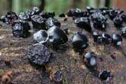 Kit Scates Barnhart |
401b Other shapes or surfaces, may be clustered or confluent, growing on hardwood or conifers, (basidia in hymenium)
................................................................................402
402a Reddish black to olive-black or black; cushion-shaped to irregularly lobed, or brain-like with folds, upper surface may have papillae; often forming masses up to 50 cm long; on rotting hardwood logs, branches or dead standing trunks
................................................................................Exidia glandulosa
| BASIDIOCARP 1-2 cm wide, cushion-shaped to irregularly lobed but often fusing to form rows or masses up to 50 cm long and 0.5 to 1.5 cm thick; transparent smoky color, becoming brownish black, reddish black, olive-black, or black, and drying black; upper surface with papillae (gland-like or wart-like protuberances, use hand lens), margin sharply bounded and not attached to substrate, no stipe. HABITAT on hardwood, rarely conifer wood, spring, fall, and winter. MICROSTRUCTURES spores 10-16 x 4-5 um, curved-cylindrical, basidia longitudinally cruciate-septate, 4-spored; clamp connections present. REMARKS Although "warts" are typical of Exidia glandulosa, they are lacking from a taxon similar to this species on shore pine and other conifers (R. Bandoni, pers. comm.). Exidia glandulosa appears to be scarce in the Pacific Northwest, at least in typical form. Exidia glandulosa forma populi differs primarily in macroscopic characters (according to Klett (1964) who regards it provisionally as a separate species): it has the inferior surface appressed but free (as opposed to adherent to the substrate) and grayish to grayish granulose, and this form lacks surface papillae (it is smooth to finely asperulate and if occasional wart-like structures are found, they are composed of crystals). Forma populi is somewhat more common in the Pacific Northwest than the typical form, particularly on Populus trichocarpa. Exidia recisa has lighter colors than Exidia glandulosa, is more erect and less inclined to fuse, has a very short stipe-like base, and lacks the warty surface. | Exidia glandulosa.jpg) Michael Wood (MykoWeb) |
402b Other colors or shapes
................................................................................403
403a 2.5-20 cm wide or more, typically a roundish mass of wavy or leaf-like folds, lobes, and convolutions; reddish cinnamon, brown, vinaceous brown, purplish brown, or (especially on drying) blackish brown; surface smooth
................................................................................Tremella foliacea
(See 614a.)
403b Not usually exceeding 5 cm, other shapes or colors
................................................................................404
404a Surface form somewhat resembling a mulberry or blackberry; dark reddish brown to almost black, discoloring the surrounding barked hardwood it grows on
................................................................................Tremella moriformis
(See 208a.)
404b Cushion-shaped, tuberculate (with small bumps); at first colorless, then dingy cinnamon to raisin color or washed with blackish
................................................................................Tremella subanomala group
(See 617a.)
COLORLESS TO WHITE OR GRAY
501a Whitish flabby, rubber-like basidiocarp 2-6 cm high that is tongue-shaped to spoon-shaped and has soft teeth on underside of the expanded upper part, common on wood and litter of conifers
................................................................................Pseudohydnum gelatinosum
| BASIDIOCARP 2-6 cm high, flabby, rubber-like basidiocarp with a head that is flattened and expanded laterally (tongue-shaped, spoon-shaped, fan-shaped, or bracket-like), attached to wood or litter by lateral stipe-like structure; usually hyaline to white, more rarely gray to gray-brown; upper (sterile) surface slightly scabrous, lower (hymenial) surface densely spiny, spines up to 3(5) mm long. HABITAT on conifer wood or litter. MICROSTRUCTURES spores 5-7 x 4.5-7 um, globose to subglobose, basidia longitudinally septate, 4-spored; cystidia not seen; clamp connections present. | Pseudohydnum gelatinosum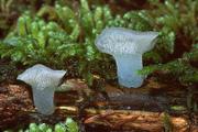 Michael Beug |
501b Shape or size different
................................................................................502
502a Basidiocarps on or associated with recognizable fungal host (basidiocarps grow on host tissue but host may be present only as a core in the basidiocarp)
................................................................................503
502b Basidiocarps on wood or on a fungal host that is not recognizable
................................................................................505
503a Basidiocarps associated with Aleurodiscus grantii
................................................................................Tremella mycetophiloides
BASIDIOCARP at first resembling liquid droplets, enlarging, the individual basidiocarps 0.1-1.5 mm in diameter, typically gregarious, often anastomosing, extending up to 1.5 cm, mostly hemispherical or somewhat disc-shaped, smooth to slightly wrinkled or brain-like; at first colorless to milky, milky to very pale pink or yellow when young. HABITAT on the hymenial surface of Aleurodiscus grantii (and elsewhere on Aleurodiscus amorphus), which predominantly occur on dead branches in live trees of Abies spp. (true fir) and infrequently on species of other conifers. The Tremella may be found on the hymenium of the Aleurodiscus basidiomes (the surface away from the wood) as minute, brownish, resin-like spots, often indented, if the material is dry. If the weather is wet, the Tremella will appear as drops of milky fluid on the surface of the Aleurodiscus. MICROSTRUCTURES basidiospores (7.0)8.0-12.5 x (7.0)8.0-10.0 um when present, subglobose to ovate or bulliform, conidia regularly present, intermixed with basidia or often alone in the hymenium, 5.0-9.5 x 3.0-5.0(6.5) um, variable, subglobose, ellipsoid, teardrop-shaped, or subcylindrical, sometimes peanut-shaped; basidia 4-spored; clamp connections abundant. REMARKS Tremella mycophaga G.W. Martin is treated here as a synonym, but not all authors agree. Tremella simplex looks similar to T. mycetophiloides and is found in eastern North America and Europe: both can occur on Aleurodiscus amorphus (as well as A. thujae for T. simplex and A. grantii for T. mycetophiloides), but T. simplex differs in having 2-celled basidia and in lacking clamp connections. DNA evidence suggests T. simplex and T. mycophaga are the genetically identical (R. Bandoni, pers. comm.).
503b Basidiocarps associated with Stereum sanguinolentum (on conifer wood)
................................................................................Tremella encephala
(See 613a.)
503c Basidiocarps associated with Peniophora spp.
................................................................................504
503d Basidiocarps associated with pyrenomycetes, (spores globose or subglobose)
................................................................................507
504a Basidiocarps 0.2-0.5 cm wide, cushion-shaped, hemispheric, discoid, infrequently anastomosing; cream to pale yellow or dull orange when moist, the interior often brownish; uncommon
................................................................................Tremella versicolor
(See 113a.)
504b Basidiocarps reaching larger sizes, becoming lobed or brain-like; often tinged yellow to orange to brown especially when young or exposed to light
................................................................................pale forms of Tremella mesenterica
(See 121a.)
505a (502b) Basidiocarps with whitish fleshy core surrounded by a gelatinous layer that is usually pale tan to pale brownish or pinkish brown but varies to yellowish, pinkish, white, or colorless; (spores 8-11 x 7-9 um)
................................................................................Tremella encephala
(See 613a.)
505b Basidiocarps without white fleshy core
................................................................................506
506a Basidiocarps quite small, usually 1-6 mm in diameter, becoming somewhat larger only by confluence, bursting out from the cavities of pyrenomycetes on wood, (spores globose to subglobose)
................................................................................507
506b Basidiocarps larger, or forming extensive masses up to 10 cm across, growing on wood but not on pyrenomycetes
................................................................................508
507a (506a, 503d) Basidiocarps milky to greenish; spores (7) 9-12 x 7.5-11 um, basidia with stalk-like base
................................................................................Tremella exigua group
(See 301b.)
507b Basidiocarps milky to light yellowish brown, may be green tinted from algae; spores 6-7.5 x 5-7 um, basidia with stalk-like base
................................................................................Tremella globispora
BASIDIOCARP 2-5 mm in diameter, up to 2(5) mm high, arising as dots from the openings made in the bark by the pyrenomycetes, becoming irregularly hemispheric cushion-shaped, by confluence reaching 10 or even 20 mm, hanging from undersides of small twigs as transparent-watery droplets, surface marked with brain-like markings or wrinkles or convolutions; pale dingy translucent whitish and opalescent, to pinkish white, cream, yellow, or light yellowish brown, sometimes discolored greenish by algae, drying to an inconspicuous film. HABITAT Tremella globispora is reported on hardwood and conifer wood, but is mycoparasitic on Diaporthe and other pyrenomycetes. Microscopically, each basidiocarp originates within a young perithecium, growing up and out of the ostiole, and expanding at the surface of the substrate. MICROSTRUCTURES (5)6.2-8.2(10.2) x 5.75-7.2(9.75) um (from deposit, excluding the well-developed apiculus), subglobose, one-celled; basidia "cruciate-septate" with oblique septa, 4-spored; hyphidia absent; clamp connections present; perithecia can be seen in early stages of development of the Tremella sporocarp.
508a (506b) Whitish or grayish or brownish gray or light yellowish brown, or dark brown with olive to violaceous tints; starting as pustules and becoming smooth or convoluted, coalescing to form irregular areas up to 10 cm across; waxy-gelatinous or gelatinous; (spores cylindrical or curved cylindrical)
................................................................................509
508b Basidiocarps often with yellow or orange tinges especially when young or exposed to light; individual basidiocarps may be several cm tall and wide, typically consisting of convoluted or brain-like lobes or folds but sometimes more shapeless, often coalescing forming irregular areas up to 10 cm across; gelatinous; (spores broadly ellipsoid)
................................................................................pale forms of Tremella mesenterica
(See 121a.)
509a Becoming spherical, brain-like or somewhat leaf-like, whitish, may turn amethyst-color or vinaceous brown, may develop numerous seed-like concretions embedded in the jelly, (spores 10-11 x 4-4.5 um, myxarioid basidia)
................................................................................Myxarium atratum
| BASIDIOCARP starts as pustules bursting through surface of substrate, becoming spherical, brain-like, or occasionally somewhat leaf-like, soon anastomosing and becoming broadly spread out up to 10 cm or more; colorless or whitish, (sometimes becoming amethyst colored or vinaceous brown but these changes lacking in reports from BC), and may have numerous seed-like 0.2-0.5 mm calcareous concretions embedded in the jelly (but these tend to be absent in Pacific Northwest collections), drying to a dark film with any concretions becoming more prominent. HABITAT on hardwood. MICROSTRUCTURES spores 10-11 x 4-4.5 um, curved-cylindrical; basidia "cruciate-septate" and myxarioid (having a stem-like part separated by a wall from the spherical metabasidial part). | Myxarium atratum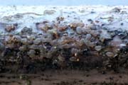 A and O Ceska |
509b Without seed-like concretions embedded in the jelly and not turning amethyst-color or vinaceous brown, (spores longer or wider or not examined, basidia not myxarioid or not examined)
................................................................................510
510a At first grayish lilac, becoming brownish gray to dark brown with olive to violaceous tints, when slightly dry appearing blackish brown; 5-25 mm across, with central point of attachment, remaining discrete disc-shaped to top-shaped, or anastomosing to form convoluted masses up to 80 mm long; surface usually roughened by conic papillae, (spores (14.5)16-19(21) x 5-6 um)
................................................................................Exidia zelleri
(See 208b.)
510b Differently colored or shaped
................................................................................511
511a Surface becoming smooth or convoluted or brain-like; color almost colorless, dingy translucent, dingy white, or pale yellow-brown, then when fully developed with a greenish or olivaceous tint; consistency waxy-gelatinous; margin abrupt; (spores 9.3-13(15.4) x 5-8.7 um, basidia not myxarioid, gloeocystidia present and becoming yellowish granular but not projecting)
................................................................................Sebacina lactescens
(This is often known as Ductifera sucina. See 301a for description.)
511b Surface undulate to alveolate to brain-like, occasionally lobed, with or without light colored or whitish papillae up to 0.5 um long; color grayish to whitish to cream to grayish orange to cinnamon to vinaceous russet, very often with more than one of these colors on one basidiocarp; consistency tough-gelatinous; margin free, undulate to incised, often fringed by long papillae; (spores 11-13.5-16 x 4-5(5.5) um, basidia not myxarioid, gloeocystidia absent)
................................................................................Exidia candida
| BASIDIOCARP appressed - disc-shaped or almost immediately anastomosing to form expanded masses up to 10 cm or more, most often broadly attached; surface undulate to alveolate to brain-like, occasionally lobed, with or without light colored or whitish papillae; undersurface appressed to free, margin free, undulate to incised, often fringed by long papillae; consistency tough-gelatinous; grayish to whitish to cream to grayish orange to cinnamon to vinaceous russet, very often with more than one of these colors on one basidiocarp. HABITAT on hardwood, fruiting time including winter and early spring. MICROSTRUCTURES spores 11-16 x 4-5(5.5) um, curved-cylindrical; hypobasidia ellipsoid to obovate to obpyriform, often with a stalk, basidia 2-4-celled by longitudinal to oblique septa; hyphidia present, apically few-branched to much-branched, occasionally unbranched; clamp connections present. REMARKS Non-papillate, unicolored collections of Exidia candida are often difficult to separate from E. repanda. The basidiocarps of E. candida are usually broadly attached, while those of E. repanda have a narrow, short, stipe-like point of attachment. Spores of E. repanda are generally narrower and hypobasidia are ellipsoid without a stalk. | Exidia candida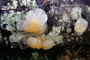 Michael Beug |
511c Surface spherical, brain-like or somewhat leaf-like, color whitish, spores 10-11 x 4-4.5 um, myxarioid basidia
................................................................................Myxarium atratum
(See 509a.)
BROWN
601a Shelving and somewhat ear-like form or inverted cup-shaped with thin rubbery (cartilaginous) flesh, the convex upper surface dark brown to reddish brown, hairy, and lower concave surface red brown to yellowish brown, hairless, and irregularly ribbed and veined
................................................................................Auricularia americana group
| BASIDIOCARP 2-10 cm, shelving and somewhat ear-like form or inverted cup-shaped, laterally or less often centrally attached, convex upper sterile surface with dense silky covering or minutely hairy, not zoned, concave lower fertile surface moist, hairless, both surfaces often wrinkled; consistency tough, flabby-gelatinous; color red-brown, cinnamon, yellow-brown, olive-brown or blackish brown; drying nearly black. HABITAT hardwoods and conifers, spring to fall. MICROSTRUCTURES spores 12-19 x 4-8 um, curved-cylindrical; basidia with 3 transverse septa, 3 lateral and 1 terminal epibasidia. REMARKS The species in the group as considered here correspond to the phylogenetic Auricularia auricula-judae complex. Members of this group include at least two North American members, Auricularia americana and Auricularia angiospermarum. The former grows on conifer wood and the latter on hardwood. Similarly colored ascomycete cups are brittle, usually terrestrial, and have their hymenium on the upper surface. Cytidia patelliformis could key out here but is very thin-fleshed and not rubbery (see 207b). | Auricularia americana group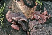 Kit Scates Barnhart |
601b Differently colored or shaped
................................................................................602
602a Whitish to gray brown; flabby, rubber-like basidiocarp 2-6 cm high that is tongue-shaped to spoon-shaped and has soft teeth on underside of the expanded upper part; common on wood and litter of conifers
................................................................................Pseudohydnum gelatinosum
(See 501a.)
602b Differently colored or shaped
................................................................................603
603a Pale to deep pink to reddish orange or salmon-colored; tongue-shaped to somewhat funnel-shaped, usually indented or split on one side, with a head and stipe, 2-8(18) cm high, higher than wide; on conifer wood, sometimes on buried twigs along streams, or on ground under conifers
................................................................................Guepinia helvelloides
(See 201a.)
603b Differently colored or shaped
................................................................................604
604a Growing as a thin film on the surface of tumor-like growths of the caps and stipes of Gymnopus dryophilus
................................................................................Syzygospora effibulata
BASIDIOCARP a thin film on the surface of galls (tumor-like outgrowths) on the surface of Gymnopus dryophilus caps and stipes. HABITAT on Gymnopus dryophilus caps and stipes. MICROSTRUCTURES spores (5.5)6-8(10) x 1.8-2.5 um, cylindrical to narrowly obovate; conidia of two types, some subglobose to broadly ellipsoid, 2-3.5 x 1.5-2 um, others rather few, cylindrical to narrowly ellipsoid, up to 13 x 2 um; basidia 2(3)-spored; clamp connections not observed. REMARKS Syzygospora mycetophila, an eastern species that also grows on the surface of galls on Gymnopus dryophilus, is indistinguishable macroscopically but has 4-spored basidia, clamp connections, and more ellipsoid spores. Two species of Syzygospora grow in BC on lichens, Syzygospora bachmanniii on Cladonia (waxy to gelatinous, reddish brown or brown, elongate to roundish or irregular, up to 5 mm long), and Syzygospora physciacearum (also found in Oregon) on Heterodermia, Physcia, and Physconia (waxy to gelatinous, pinkish, pale brown, reddish brown, grayish brown to almost black, convex, often with a constricted base, up to 1.2 mm long).
604b Differently shaped
................................................................................605
605a Blackish on upper surface, top-shaped to shallow cup-shaped, with considerable gelatinous material beneath the hymenium, smooth on hymenium, dark brown and roughened on exterior, clustered but not confluent, growing on hardwood; (spores spores 9-17 x 6-7.5 um, 4 dark spores and 4 lighter ones in each ascus in hymenium)
................................................................................Bulgaria inquinans
(See 401a.)
605b Purple brown, saucer-shaped to rounded to lobed or brain-like, smooth, growing on hardwood, (ascospores 6-10 x 3.5-5 um)
................................................................................Ascotremella faginea
| ASCOCARP 2-4 cm across, saucer-shaped to rounded to lobed or brain-like, sometimes forming masses up to 10 cm or more; pinkish when young, becoming pinkish brown to violet brown, surface smooth, shiny to dull, short central stem-like part for attachment. HABITAT on branches and trunks of hardwoods lying on the ground, including beech and alder, summer to fall. MICROSTRUCTURES spores 6-10 x 3.5-5 um, ellipsoid, often with truncate ends, colorless, smooth except for 3-4 striations which are difficult to see (oil immersion), two droplets; asci 70-90 microns, iodine negative. REMARKS The illustrated collection from Whidbey Island in November is either this species, found in eastern North America and elsewhere, or Neobulgaria pura var. foliacea. Differentation by the lack of longitudinal striations on mature spores and the positive iodine reaction on ascal pore was not possible. The distinction may be moot, because Baral (unpublished comments on Fungi of Switzerland) says that the type of the latter seems to be a mixture of Neobulgaria pura var. pura (with a different form) and Ascotremella faginea. | Ascotremella faginea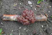 Ian Gibson |
605c Differently shaped or colored, growing on hardwood or conifer wood, (basidia in hymenium)
................................................................................606
606a Variously formed, spread out flat and irregularly rounded, brain-like, mesentery-like, or weakly cup-shaped; ocher-pink, grayish-ocher to salmon-colored; on shredded inner bark of Populus trichocarpa (or on Prunus spp.)
................................................................................Craterocolla cerasi
(See 203a.)
606b Differently shaped or colored
................................................................................607
607a Orange brown or yellow brown in parts, but generally showing orange or yellow in some parts when fresh
................................................................................101
607b Differently colored
................................................................................608
608a Reddish black to olive-black or black; cushion-shaped to irregularly lobed, or brain-like with folds, upper surface may have papillae; often forming masses up to 50 cm long; on rotting hardwood logs, branches or dead standing trunks
................................................................................Exidia glandulosa
(See 402a.)
608b Differently colored or shaped
................................................................................609
609a Amber to dark red-brown or liver brown; mostly obconic when separate, forming clusters with little anastomosis, may be plate-shaped, conchate, wavy, brain-like, and/or lobed, basidiocarps up to 20 mm high, but usually about 5 mm high, 5-30 mm across, gregarious but usually with separation from other basidiocarps, central attachment almost like a stipe
................................................................................Exidia recisa
BASIDIOCARP up to 3 cm in diameter, up to 1.5 cm high, mostly obconic when separate, forming clusters with little anastomosis, may be plate-shaped, conchate, wavy, brain-like, and/or lobed, centrally attached by short stipe-like base; amber to yellowish brown, cinnamon brown, pale liver-brown or dark red-brown, drying blackish. HABITAT on dead limbs of hardwoods, particularly Salix (willow), spring to fall. MICROSTRUCTURES spores 9-14 x 3-5 um, curved-cylindrical; basidia 4-spored, longitudinally "cruciately septate"; colorless dendrohyphidia; clamp connections present; also conidia 5-6 x 1.5-2 um. REMARKS Exidia glandulosa lacks a stipe and is darker in color. Exidia repanda is disc-shaped to cup-shaped. This may be the commonest Exidia species in the Pacific Northwest if its name is correct. Klett (1964) designates Bandoni 945 and 2743 from BC (the latter deposited at University of British Columbia as E. recisa), as well as six of his own collections from Washington as Exidia crenata (Schw.) Fr., but says that he cannot say whether the latter species is the same as Exidia recisa of Europe. A somewhat similar taxon occurs on spruce and pine wood. Klett (1964) provisionally identifies this as Exidia pinicola (Peck) Coker. The collections he assigns here had mostly been identified as Exidia saccharina, or in two cases as Exidia umbrinella. Basidiocarps are at first pustulate, about 4 mm, becoming disc-shaped with a single point of attachment, then broadly expanded and appressed, anastomosing into large clusters that are ridged to brain-like, smooth or occasionally bearing crystalline warts. Color is light watery yellow at first becoming grayish orange to reddish brown. Spores are 10-15 x (3.5)4-5 um, curved-cylindrical to bent-cylindrical, basidia 2-4 celled by longitudinal septa, and clamp connections conspicuous.
609b Differently colored or shaped
................................................................................610
610a Usually growing on dead birch wood, sometimes other hardwoods; brownish translucent, pinkish brown, cinnamon brown, gray brown, olivaceous brown, or yellowish brown; basidiocarps thick button-like, the margin abrupt and rounded, up to 20 mm across, remaining separate or sometimes anastomosing but a contact line remaining when anastomosing, at first pustulate, then disc-shaped to cup-shaped, surface flat to undulating or wrinkled, occasionally with sparse fine warts, centrally attached by a point
................................................................................Exidia repanda
BASIDIOCARP up to 2.5 cm broad, disc-shaped (like a thick button in form), becoming cup-like, the margin abrupt and rounded, basidiocarp usually pressed down close to the wood but only centrally attached, not readily anastomosing, if anastomosing a contact line remaining, smooth to furrowed or wrinkled, sometimes with sparse fine warts, underside smooth, margins thick; pale brown to yellowish brown, pinkish brown, cinnamon brown, grayish brown, or olivaceous brown, drying dark brown; underside the same color or slightly lighter. HABITAT dead birch and sometimes other hardwood. MICROSTRUCTURES spores mostly 11-15 x 3-4 um, curved-cylindrical; basidia "cruciate-septate" or sometimes merely 2-celled; hyphidia present; clamp connections present. REMARKS Exidia zelleri may have similar form but color is different (see 611a).
610b Differently colored or shaped
................................................................................611
611a At first grayish lilac, becoming brownish gray to dark brown with olive to violaceous tints, when slightly dry appearing blackish brown; 5-25 mm across, with central point of attachment, remaining discrete disc-shaped to top-shaped, or anastomosing to form convoluted masses up to 80 mm long; surface usually roughened by conic papillae
................................................................................Exidia zelleri
(See 208b.)
611b Differently colored or shaped
................................................................................612
612a Grayish to whitish to cream to grayish orange to cinnamon to vinaceous russet, very often with more than one of these colors on one basidiocarp; arising as colorless pustules, expanding to appressed-discoid or quickly anastomosing to form masses 10 cm or more across, most often broadly attached; surface undulate to alveolate to brain-like, with or without light colored or whitish papillae; margin undulate to incised, often fringed by papillae
................................................................................Exidia zelleri
(See 511b.)
612b Differently colored or shaped
................................................................................613
613a Basidiocarps with whitish fleshy core surrounded by a gelatinous layer that varies from pale pinkish brown, yellowish brown, or pale tan, to white or colorless; up to 60 mm but usually 3-10 mm across and up to 5 mm high, irregularly hemispheric - cushion-shaped, surface bumpy-undulating-furrowed, may be brain-like; (spores 8-11 x 7-9 um)
................................................................................Tremella encephala
| BASIDIOCARP irregularly hemispheric - cushion-shaped, appressed flat on substrate, upper spore-bearing surface bumpy-undulating and furrowed or pleated-wrinkled, sometimes brain-like, margin sharply bounded, may be 0.3-1.0 cm across and up to 0.5 cm high, but often larger reaching 6 cm across, basidiocarps in section through the center of the base with leathery-fleshy white core surrounded by gelatinized material, subsessile; varies from pale pinkish brown, yellowish brown, or pale tan, to pinkish, yellowish, white or colorless, drying brown. HABITAT Basidiomes are composed of a dense core of Stereum sanguinolentum hyphae, surrounded by a thin layer of Tremella hyphae, hymenium, etc., but typically reported on dead coniferous wood, less commonly hardwood. Occasional small basidiocarps occur directly on the lobes of the closely situated Stereum. MICROSTRUCTURES spores 8-11 x 7-9 um, globose to short-ellipsoid; basidia, longitudinally septate, 2-spored or 4-spored; clamp connections present. REMARKS The core of the basidiocarp consists of transformed hyphae of Stereum sanguinolentum. This is the only large Tremella that occurs on conifer wood. A species resembling this on Prunus has been found to differ in DNA studies (R. Bandoni, pers. comm.) | Tremella encephala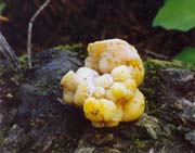 Buck McAdoo |
613b Basidiocarps without white fleshy core
................................................................................614
614a Reddish cinnamon, brown, vinaceous brown, purplish brown, or blackish brown; 2.5-20 cm wide or more, typically a roundish mass of wavy or leaf-like folds, lobes, and convolutions; surface smooth; on dead hardwood or conifer wood, bleeding Stereum species may be nearby
................................................................................Tremella foliacea
| BASIDIOCARP 2.5-20 cm wide or more, typically a somewhat spherical mass of wavy or leaf-like folds, lobes, and convolutions; stipe absent but growing together from a common base; reddish-cinnamon, brown, vinaceous brown, purplish brown, or blackish brown, drying blackish brown. HABITAT on dead wood of hardwoods or conifers, associated with bleeding Stereum species (the color possibly derived from the host fungus). MICROSTRUCTURES spores 8-9(13) x 7-9 um, ovate to globose; basidia longitudinally septate, 2-4-spored; hymenial conidia absent; clamp connections present. REMARKS "Like the T. aurantia group, T. foliacea and its allies are parasites of Stereum spp. and of at least one morphologically similar but unrelated form. Sporocarps of the T. foliacea group contain hyphae of the Stereum or other host, but these hyphae are not in the proportions seen in T. encephala and T. aurantia. They are generally visible only in microscope preparations of the sporocarp lobes." (R. Bandoni, pers. comm.) | Tremella foliacea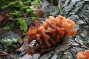 A and O Ceska |
614b Differently colored or shaped
................................................................................615
615a Dark reddish brown to almost black, discoloring the surrounding barked hardwood it grows on; somewhat resembling a mulberry or blackberry
................................................................................Tremella moriformis
(See 208a.)
615b Differently colored or shaped
................................................................................616
616a Buff to brown, maroon, red, flame-colored, orange, or yellow, sometimes punctate with darker brown spots; up to 50 mm long and 13 mm high, often begins as a lobe protruding through ruptured bark, developing leaf-like lobes or becoming brain-like or top-shaped; growing on dead attached branches of dogwood and willow; often associated with Peniophora basidiocarps
................................................................................Tremella mesenterella
(See 121b.)
616b Differently colored or shaped
................................................................................617
617a Basidiocarps at first colorless, then dingy cinnamon to raisin color or washed with blackish; cushion-shaped, surface with small bumps; (spores 8-10.5(14) x 10-11(14) um, often wider than long)
................................................................................Tremella subanomala - T. indecorata group
BASIDIOCARP up to 4 cm in longest dimension, convex, cushion-shaped; at first colorless, then dingy cinnamon to raisin color or washed with blackish, drying blackish cinnamon or fuscous. HABITAT on dead hardwood, likely associated with pyrenomycete fungi. MICROSTRUCTURES spores 8-10.5(14) x 10-11(14) um, subglobose, often wider than long; basidia becoming "cruciate-septate". REMARKS Tremella subanomala has been reported in the Pacific Northwest from Oregon and British Columbia, but is closely related to or possibly the same as T. indecorata Sommerf. T. albida Huds.: Fr. is also closely related. Bourdot and Galzin (1928) discuss several varieties of T. indecorata; similar forms occur in British Columbia and need intensive study to determine the number of taxa.
617b Differently colored or shaped
................................................................................618
618a Basidiocarps milky to light yellowish brown, may be green tinted (when very old) from algae; 4-5 mm in diameter, reaching 10 mm by confluence, often hanging from small twigs as transparent-watery droplets, surface with brain-like markings or wrinkles, arising as dots from the openings made in the bark by the pyrenomycetes; (spores 6-7.5 x 5-7 um, basidia with stalk-like base)
................................................................................Tremella globispora
(See 507b.)
618b Whitish or grayish basidiocarp starting as pustules and becoming smooth or convoluted, coalescing to form irregular areas up to 100 mm across, waxy-gelatinous or gelatinous; (spores cylindrical or curved cylindrical)
................................................................................619
Note: A reddish brown colored Cytidia patelliformis might key out here but it is thin-fleshed, resupinate to open cup-shaped (see 207b).
619a Colorless or whitish becoming vinaceous brown; pustules bursting through surface of bark or wood, becoming brain-like or somewhat leaf-like, soon anastomosing and up to 100 mm or more across; sometimes developing numerous seed-like concretions embedded in the jelly; (spores 10-11 x 4-4.5 um, myxarioid basidia, gloeocystidia not present)
................................................................................Myxarium atratum
(See 509a.)
619b White or gray to light ochraceous, not turning vinaceous brown; without seed-like concretions embedded in the jelly; (spores 9.3-13(15.4) x 5-8.7 um, basidia not myxarioid, gloeocystidia present and becoming yellowish granular but not projecting)
................................................................................Sebacina lactescens
(This is often known as Ductifera sucina. (See 301a for description.)
adnexed - lightly attached
alveolate - surface of structure with broad pits
angiosperm - plant producing flowers and reproducing by seed produced in a carpel, including most flowering plants and trees but not conifers which are gymnosperms
arthoconidium - arthrospore, conidium converted from an existing, determinate hyphal element
arthrospore - same as arthroconidium, or a specialized uninucleate cell functioning as a spore and derived from the disarticulation of cells of a formerly vegetative branch (Kirk et al.)
ascocarp - ascus-bearing fruiting body
ascoma (plural ascomata) - ascus-bearing fruiting body
ascospore - spore formed in an ascus by a species of the ascomycetes
ascus (plural asci) - typically sac-like cell characteristic of Ascomycota in which ascospores (most often 8) are produced
aseptate - without a septum
asperulate - roughened with small warts or points
basidiocarp - basidium-bearing fruiting body
basidiome - basidium-bearing fruiting body
basidiospore - spore borne on a basidium
basidium (plural basidia) - This is the cell on which spores form in species of basidiomycetes. More specifically, the basidium is the cell or organ from which after meiosis, basidiospores (most often 4) are produced externally, each on an extension called a sterigma. The morphological part or developmental stage of the basidium in which karyogamy occurs is called the probasidium. The morphological part or developmental stage of the basidium in which meiosis occurs after that is called the metabasidium. The terms hypobasidium and epibasidium are best avoided. A holobasidium is a basidium in which the metabasidium is not divided by primary septa. A homobasidium is a basidium of a Homobasidiomycete, usually a holobasidium. A phragmobasidium is a basidium in which the metabasidium is divided by primary septa, usually "cruciate-septate" (e.g. Tremella, but see entry for that term) or transverse (e.g. Auricularia). A heterobasidium is a basidium of a Heterobasidiomycete, usually a phragmobasidium. In Dacrymycetales (e.g. Dacrymyces), the forked metabasidium having 2 sterigmata each bearing a spore is characteristic.
bullate - of a surface, with knob-like or blister-like (bubble-like) projection(s), sometimes used to referred to bulliform spores
bulliform - of a spore, circular in top to bottom view and elliptic in side view: in the side view the apiculus more or less centrally placed on one of the longer sides; the word "bulliform" is derived from Latin "bulla" meaning a "knob", and the spores could be regarded as "doorknob-shaped", (R. Bandoni, pers. comm.)
cerebriform - brain-like in shape
cespitose - growing in tufts or close clusters from a common base, but not grown together
conchate - shell-shaped, variously held to be like a conch, like a bivalve shell, or like an oyster shell
conidium (plural conidia) - a non-motile, thin-walled, asexual spore, usually falling off readily, not developed by cytoplasmic cleavage
conidiogenous - producing conidia
conidioma (plural conidiomata) - conidium-bearing multi-hyphal structure or fruiting body
cruciate-septate - of basidial septa (in Tremellales), vertical (longitudinal) and at right angles. (This term is an inaccurate one in that the septa do not actually "cross" one another.- as can plainly be seen in species where the basidial cells often separate a bit if the basidiome is dried and revived several times. Each of the 4 cells has a separate wall.)
decorticate - without bark
deliquescence - the act or process of melting or dissolving into liquid
dendrohyphidium (plural dendrohyphidia) - branched, slender, sterile element in hymenium or subhymenium
dicotyledonous - of angiosperms, having two embryonic leaves as opposed to monocotyledonous angiosperms which have one
dikaryophysis (plural dikaryophyses) - modified terminal hypha in the hymenium, now generally replaced by hyphidium: synonyms or near synonyms include paraphysis, paraphysoid, pseudoparaphysis, pseudophysis, hyphidium and dikaryoparaphysis, but the term is particularly used when describing jelly fungi (e.g. Tremellales, Dacrymycetales), see hyphidium for more details on these terms.
discoid - dish-shaped, usually with a central attachment; flat and circular, resembling a disc
embedded - of hymenial elements such as cystidia, neither reaching the surface formed by other elements (such as basidia) nor projecting beyond it
epibasidium (plural epibasidia)- In the context of jelly fungi, the epibasidium is the part of the basidium closest to the sterigma, but the term has been used in different senses and is best avoided where possible. In the terminology of Neuhoff (1924), it is the name given to each of the 4 appendages that are separated from the basal cell of hypobasidium by a septum, after this appendage has received its cytoplasm and nucleus, and each of which bears a sterigma and spore, the hypobasidium and epibasidium making up a heterobasidium. G.W. Martin (1937) applies the term to any structure that intervenes between the hypobasidium and a sterigma (or rarely to an asterigmate spore). G.W. Martin (1957) uses the term in a somewhat different way.
erumpent - sporocarp erupting through its substrate but barely rising above it
foliaceous - resembling a leaf or leaves
fusiform - spindle-shaped, fairly slender and narrowing from middle to both ends
gloeocystidium (plural gloeocystidia) - thin-walled cystidium, usually irregular, contents colorless or yellowish and highly refractile, (Kirk et al.)
haustorial - pertaining to haustoria, which are special hyphal branches, especially one within the living cell of the host, for absorption of food, (Kirk et al.)
heterobasidiomycete - see basidium and Tremellomycetidae
holobasidium (plural holobasidium) - see basidium
homobasidiomycete - see basidium and Tremellomycetidae
hyaline - colorless, or at least translucent if faintly colored
hymenium - fertile area of sporocarp where spores are produced
hyphidium (plural hyphidia) - the preferred name for a modified sterile terminal hypha in the hymenium: synonyms or near synonyms include dikaryophysis (often used for jelly fungi such as Tremellales, Dacrymycetales: the name refers to the fact that the hyphidia are dikaryotic), paraphysis (used particularly for ascomycetes, derived from haploid hyphae), pseudoparaphysis (same as hyphidium for basidiomycetes, but a type of hamathecium in ascomycetes hamathecium being a neutral term for hyphae or other tissues between asci or projecting into the locule or ostiole of ascomata, different types of hamathecium include paraphysis, paraphysoid, pseudoparaphysis, periphysoid, and periphysis)
hymenophore - spore-bearing surface or structure
indeterminate - having the edge or shape not well defined
lamellate - having lamellae (gills); made up of thin plates, (Kirk et al.)
muriform - of spores, "having cells arranged like bricks in a wall, with both longitudinal and transverse septa", (Snell & Dick)
mycoparasitic - if one fungus (a mycoparasite) is parasitic on another fungus it is said to be mycoparasitic
mycorrhiza (plural of mycorrhizae) - a particular symbiotic relationship with the roots of a seed plant, wherein the rootlets of trees are covered or permeated by the mycelium of fungi
myxarioid - of basidia, having a stem-like part separated by a wall from the spherical metabasidial part, as in Myxarium, (Kirk et al.)
obcone - a cone with the point down
obconic - like an ice-cream cone with point down
obovoid - ovoid but with the larger end in the opposite direction to the usual
obovate - ovate with the larger end in the opposite direction to the usual
obovoid - ovoid with the larger end in the opposite direction to the usual
obpyriform - pear-shaped in the opposite direction to the usual one
ostiole - the opening at the top of many fungal sporocarps (perithecia, pycnidia, puffballs) through which spores escape or are expelled
ovate - similar to ovoid but some regard as more pointed at the narrower end
ovoid - like the shape of an egg
palmate - "having lobes radiating from a common centre but not extending to the point of insertion", (Kirk et al.)
paraphysis (plural paraphyses) - modified terminal hypha in the hymenium, synonyms or near synonyms include paraphysoid, pseudoparaphysis, pseudophysis, hyphidium and dikaryoparaphysis, (Kirk et al.), see hyphidium for more details
pedicel - a small stalk or stalk-like attachment
perithecium (plural perithecia) - a nearly spherical, ovoid, pear-shaped or beaked (flask-like) body bearing asci in the interior, with or without sterile paraphyses, with an ostiole (opening), characteristic of the pyrenomycetes
poroid - with pores or structures that look like pores
probasidium (plural probasidia) - see basidium
pruinose - looking finely powdered or finely granular
pulvinate - cushion-shaped, shaped like a small circular cushion and usually distinctly convex
punctate - marked with dots consisting of hollows, depressions, spots, raised-joined scales, or agglutinated fibrils, all very small
pustulate - having pustules
pustule - pimple-like or blister-like elevation
pyrenomycete - member of the Class Pyrenomycetes, now not generally accepted as a class, and used in various senses but mostly for fungi with perithecia that bear unitunicate asci
resupinate - lying flat on what it is growing on, without a stem or well-formed head, with the spore-bearing structures facing outward
scabrid - rough with delicate and irregular projections (Kirk et al.)
scurfy - surface covered with bran-like particles resembling scales, same as furfuraceous
sessile - lacking a stipe or stalk
spathulate - shaped like a spatula or spoon, rounded elongate with rounded or obtuse tip and with a narrowing or stalk-like base
sporocarp - a body in which or on which spores are produced, a more precise word for fruiting body
stellate - star-shaped
stipitate - having a stipe (or stem)
stroma (plural stromata) - "a mass or matrix of vegetative hyphae, with or without tissue of the host or substrate, sometimes sclerotium-like in form, in or on which spores or fruit bodies are produced", (Kirk et al.)
subcortical - under the bark; of the subcortex (of the layer below the cortex)
subglobose - nearly spherical
subpyriform - somewhat pyriform (somewhat pear-shaped), almost pyriform
subsessile - almost sessile (almost lacking a stipe or stalk)
substipitate - with a narrowed base but not a clearly differentiated stem
substrate - the material that a fungus is growing on or attached to, in this sense the same as substratum
subtending - supporting underneath
symbiont - organism living in symbiosis with another, with mutual benefit
telial - pertaining to a telium, the structure producing teliospores
teliospore - the spore (commonly a winter or resting spore) of the Uredinales (or Ustilaginales) from which the basidium is produced (Kirk et al.)
tomentose - covered with soft hairs, often soft densely matted hairs, like a woollen blanket
Tremellaceous - of or pertaining to the Tremellales, having the characteristics of this order, (Snell & Dick)
Tremellomycetidae - Tremellomycetidae is the subclass of Basidiomycetes which comprises those basidiomycetes in which the metabasidium is divided by primary septa (usually cruciate or horizontal), including Auriculariales, Ceratobasidiales, Christenseniales, Cystofilobasidiales, Dacrymycetales, Filobasidiales, Tremellales, and Tulasnellales. The other Basidiomycetes are in the subclass Agaricomycetidae, in which the metabasidium is not divided by primary septa but may sometimes become adventitiously septate. A similar group to the Tremellomycetidae is the heterobasidiomycetes which have a basidium often indefinite in form and usually septate, bearing spores that germinate to form secondary spores or to form sprout mycelium, as opposed to homobasidiomycetes which have basidia with a definite form that is non-septate, bearing spores that germinate to form true mycelium. Dacrymycetales, Tremellales, and Auriculariales are generally among those included in the heterobasidiomycetes. Phragmobasidiomycetes are another similar group which have a basidium in which septation follows directly after the first and second divisions of the primary, basidial nucleus, so that the mature basidium is divided into four cells, as opposed to holobasidiomycetes which have a basidium in which nuclear division is not followed by formation of septa. Most jelly fungi (including including Tremellales and Auriculariales) are phragmobasidiomycetes, but other basidiomycetes including Dacryomycetales are holobasidiomycetes.
tuberculate - with (usually) sparse and irregular warts; having tubercles, synonym of punctate (Kirk et al.)
urceolate - having the shape of a pitcher, with large body and small mouth
Urediniomycetidae - a subclass of Basidiomycetes which includes the rust fungi and related forms with simple septal pores
villose - covered with long soft, weak hairs that collapse readily
| GENUS AND SPECIES | KEY ENTRIES |
| ASCOCORYNE J.W. Groves and D.E. Wilson | |
| A. cylichnium (Tul.) Korf | 202a |
| A. sarcoides (Jacq.) J.W. Groves & D.E. Wilson | 202a |
| ASCOTREMELLA Seaver | |
| A. faginea (Peck) Seaver | 202b, 605b |
| AURICULARIA Bull. ex Juss. | |
| A. americana Parmasto & I. Parmasto ex Audet, Boulet & Sirard | 601a |
| BULGARIA Fr. | |
| B. inquinans (Pers.) Fr. | 401a, 605a |
| CALOCERA (Fr.) Fr. | |
| C. cornea (Batsch: Fr.) Fr. | 102a |
| C. viscosa (Fr.) Fr. | 101a |
| CRATEROCOLLA Bref. | |
| C. cerasi (Schumach.) Bref. | 203a, 206a |
| CRONARTIUM Fr. | 135b |
| CYTIDIA Quél. | |
| C. lanata W.B. Cooke | 207c |
| C. patelliformis (Burt) A.L. Welden | 207b |
| C. salicina (Fr.) Burt | 117a, 207a |
| DACRYMYCES Fr. | |
| D. aquaticus Bandoni & G.C. Hughes | 122a |
| D. capitatus Schwein. | 132b |
| D. chrysocomus (Bull. ex Fr.) Tul. | 126a |
| D. chrysospermus Berk. & M.A. Curtis | 119a, 124a |
| = Dacrymyces palmatus (Schwein.) Bres. | |
| D. minor Peck | 132a |
| D. minutus (L.S. Olive) McNabb | 129a |
| D. ovisporus Bref. | 128a |
| D. palmatus (Schwein.) Bres. | |
| See Dacrymyces chrysospermus | |
| D. stillatus Nees: Fr. | 127a, 131a |
| D. tortus (Willd.) Fr. | 130a |
| D. variisporus McNabb | 126b |
| DACRYOPINAX G.W. Martin | |
| D. spathularia (Schwein.) G.W. Martin | 109b |
| DUCTIFERA Lloyd | |
| D. sucina (Moeller) K. Wells | |
| See Sebacina lactescens | |
| EXIDIA Fr. | |
| E. candida Lloyd | 511b, 612a |
| E. glandulosa (Bull.) Fr. | 402a, 608a |
| E. glandulosa (Bull.) Fr. f. populi Neuhoff | 402a |
| E. nucleata (Schwein.) Burt | |
| See Myxarium atratum | |
| E. pinicola (Peck) Coker | 609a |
| E. recisa (Ditmar) Fr. | 133d, 609a |
| E. repanda Fr. | 610a |
| E. saccharina (Alb. & Schwein.): Fr. | 609a |
| E. umbrinella Bres. | 609a |
| E. zelleri Lloyd | 208b, 510a, 611a |
| FEMSJONIA Fr. | |
| F. pezizaeformis (Lev.) P. Karst. | 105a |
| GELATINODISCUS Kanouse & A.H. Sm. | |
| G. flavidus Kanouse & A.H. Sm. | 104a |
| GUEPINIA Fr. | |
| G. helvelloides (DC.) Fr. | 109a, 201a, 603a |
| = Phlogiotis helvelloides (DC.) G.W. Martin | |
| = Tremiscus helvelloides (DC.) Donk | |
| GUEPINIOPSIS Pat. | |
| G. buccina (Pers.: Fr.) L.L. Kenn. | 106a |
| G. alpina (Tracy & Earle) Brasf. | |
| See Heterotextus alpinus | |
| GYMNOSPORANGIUM R. Hedw. ex DC | 135a |
| HETEROTEXTUS Lloyd | |
| H. alpinus (Tracy & Earle) G.W. Martin | 107a |
| = Guepiniopsis alpina (Tracy & Earle) Brasf. | |
| H. luteus (Bres.) McNabb | 107b |
| MYXARIUM Wallr. | |
| Myxarium atratum (Peck) Ginns & Lefebvre | 509a, 511c, 619a |
| = Exidia nucleata (Schwein.) Burt | |
| PHLOGIOTIS Quél. | |
| Phlogiotis helvelloides (DC.) G.W. Martin | |
| See Guepinia helvelloides | |
| PSEUDOHYDNUM P. Karst. | |
| Pseudohydnum gelatinosum (Scop.: Fr.) P. Karst. | 501a, 602a |
| SEBACINA Tul. & C. Tul. | |
| Sebacina lactescens Burt | 301a, 511a, 619b |
| = Ductifera sucina sensu auct. | |
| SYZYGOSPORA G.W. Martin | |
| Syzygospora effibulata (Ginns & Sunhede) Ginns | 134a, 604a |
| TREMELLA Pers. | |
| Tremella aurantia Schwein.: Fr. | 112d, 120a |
| Tremella encephala Pers.: Fr. | 112e, 133a, 204a, 503b, 505a, 613a |
| Tremella exigua Desm. | 301b, 507a |
| Tremella foliacea (Pers.) Fr. | 205a, 403a, 614a |
| Tremella indecorata Sommerf. | 404b, 617a |
| Tremella globispora D.A. Reid | 112f, 133b, 507b, 618a |
| Tremella mesenterella Bandoni & Ginns | 114b, 121b, 616a |
| Tremella mesenterica Retz.: Fr.. | 114a, 121a, 504b |
| Tremella moriformis Sm. & Sowerby: Fr. | 208a, 404a, 615a |
| Tremella mycetophiloides Kobayasi | 112b, 133c, 503a |
| Tremella subanomala Coker | 404b, 617a |
| Tremella subencephala Bandoni & Ginns | 112a |
| Tremella versicolor Berk. & Broome | 113a, 504a |
| TREMISCUS (Pers.) Lév. | |
| Tremiscus helvelloides (DC.) Donk | |
| See Guepinia helvelloides |
- END -